Quantitative assessment of contractile reserve during dobutamine Doppler myocardial imaging echocardiography
Marina Deljanin Ilić, Stevan Ilić, Dragan Djordjević, Ivan Tasić, Ljubiša Nikolić, Todorka Savić
Institute for preventiontion, treatment and rehabilitation rheumatic and cardiovascular diseases, Niška Banja
Adresss:
Prof. dr Marina Deljanin
Ilić,
Institute for preventiontion, treatment and rehabilitation rheumatic
and cardiovascular diseases,
18 205 Niška Banja
Tel. 018 541 414
E mail: marinadi@bankerinter.net
The presence of dysfunctional but viable myocardium in patients with recent myocardial infarction has therapeutic implications since revascularization may improve both functional status and survival (1). Because coronary revascularization is beneficial in this situation, it is important to delineate viable from non viable dysfunctional myocardium in post-myocardial infarction patients. Several non-invasive techniques have been developed to assess myocardial viability, including positron emission tomography, single-photon emission computed tomography and magnetic resonance imaging (2). Dobutamine stress echocardiography (DSE) has also been used to study myocardial viability, particularly in low dose protocols in order to evaluate myocardial contractile reserve (3,4) in myocardial regions with resting wall motion abnormalities. Improvement of wall motion during low-dose (10 mcg/kg/min) dobutamine stress indicates viable myocardium while worsening of the wall motion pattern during high-dose (up to 40 mcg/kg/min) dobutamine stress is indicative of coronary stenosis causing ischemia. However, dobutamine echocardiography has some potential limitations, and one of them is that it is only a semiquantitative method. The new ultrasoud methodology, pulsed wave Doppler tissue sampling, can compensate conventional stress echocardiography limitations. It has the potential for quantitating myocardial velocity, and to assess regional left ventricular dynamics, including regional systolic and diastolic function (5,6).
This study evaluated the use of pulsed wave Doppler myocardial imaging (PW DMI), in combination with left ventricular stimulation, using dobutamine for the identification of contractile reserve in patients after acute myocardial infarction.
Material and methods
Doppler myocardial imaging. At baseline and after dobutamine infusion in all patients PW DMI studies were performed using apical transthoracic echocardiographic Doppler spectrum. PW DMI recording was obtained by positioning a sample volume in each of the 11 myocardial wall segments. Wall motion velocities was then detected throughout each cardiac cycle and displayed in the graphic format of a Doppler spectrum. In each adequately visualized segment the presence of post systolic motion (PSM) was analyzed and peak myocardial velocity (m.v.) of systolic (S), PSM, early (E) and late (A) diastolic waves were measured and ratio E/A was calculated. For each segment a frozen image of the PW DMI signals from three consecutive cardiac cycle was obtained at the end of datum acquisition sequence for of-line analysis. The final value represented the mean of three consecutive cardiac cycles.
Results
During LDDE ventricular arrhythmias occured in 5 (9,2%) patients and supraventricular in three patients. No patient had sustained arrhythmia, significant decreased or increased in blood pressure, worsening of wall motion or significant symptoms and none required interruption of the test.
Two-dimensional DSE. Visual assessment of wall motion was possible in 556(93,6%) out of 594 segments. A total of 359 (64,6%) demonstrated normal function, and 197(35,4%) segments were dysfunctional. Out of them 102 were hypokinetic, 84 akinetic and 11 dyskinetic. At LDDE 84 (42,6%) segments in 26 (48,1%) patients demonstrated contractile reserve, while 113 dysfunctional segments did not improve motion during LDDE.
Doppler myocardial imaging. Analysis of PW DMI samplings was feasible in 168(85,3%) of 197 dysfunctional segments, including 73 (86,9%) segments with and 95(84,1%) segments without contractile reserve, divided on the basis of the LDDE results. At baseline PW DMI examination PSM was present in all segments with contractile reserve and in 8/95 (8,4%) segments without contractile reserve. Baseline values of peak E diastolic velocity, S velocity and ratio E/A in segments with contractile reserve were significantly higher, compared to the values of the same parameters in segments without contractile reserve ( table 1).
Table 1. Baseline values of PW DMI parameters in segments with
and in segments without contractile reserve (CR)
|
Parameters |
With CR n = 73 |
Without CR n = 95 |
P |
|
E cm/s |
6.6 ± 2.4 |
5.3 ± 2.3 |
0.001 |
|
A cm/s |
7.7 ± 1.7 |
7.3 ± 1.9 |
NS |
|
E/A |
0.85 ± 0.19 |
0.73 ± 0.23 |
0.001 |
|
S cm/s |
6.2 ± 2.3 |
5.1 ± 2.4 |
0.005 |
E – peak early diastolic velocity; A – peak late diastolic velocity;
E/A ratio; S – peak systolic velocity
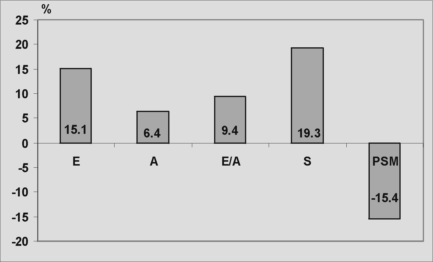
After LDDE in segments with contractile reserve, E m.v. increased by 15,1%, A m.v. increased by 6,4%, S m.v. increased by 19,3% and ratio E/A increased by 9,4%, while PSM m.v. decreased by 15,4% compared to baseline values ( figure 1 ).

Figure 1. Changes in myocardial velocities in segments with LDDE
induced contractile reserve: E – peak early diastolic velocity;
A – peak late diastolic velocity; E/A ratio; S – peak systolic velocity;
PSM – peak velocity of post-systolic motion
In segments with LDDE induced improvement of wall motion, values of peak E diastolic velocity, systolic velocity and ratio E/A were significantly higher, and value of PSM velocity significantly lower compared to the values before LDDE ( table 2 ). In segments without wall motion improvement with dobutamine, no significant changes of m.v. were detected.
Table 2. Values of PW DMI parameters before and after LDDE
in segments with and without contractile reserve (CR)
|
Parameters |
Before LDDE |
After LDDE |
P |
|
E cm/s |
6.6 ± 2.4 |
7.6 ± 2.5 |
0.02 |
|
A cm/s |
7.7 ± 1.7 |
8.2 ± 1.9 |
NS |
|
E/A |
0.85 ± 0.19 |
0.93 ± 0.21 |
0.02 |
|
S cm/s |
6.2 ± 2.3 |
7.4 ± 2.5 |
0.005 |
|
PSM cm/s |
7.1 ± 2.5 |
6.0 ± 2.3 |
0.01 |
E – peak early diastolic velocity; A – peak late diastolic velocity;
E/A ratio; S – peak systolic velocity; PSM – peak velocity of post-
systolic motion
In patients after myocardial infarction, the distinction between irreversibile fibrotic scar and dysfunctional but viable myocardium has important therapeutic and prognostic implications. A large variety of techniques have been introduced to assess viable myocardium and the most cost-effective imaging techniques to detect reversible contractile function currently are stress echocardiography and nuclear perfusion / metabolism imaging. Dobutamine stress echocardiography has been demonstrated to be a reliable technique for identification of myocardial viability (9, 10). Several protocols of dobutamine administration have been proposed (11). In the present study we used low-dose dobutamine infusion protocol because previous study have demonstrated that the monophasic response at a low-dose identifies most viable segments and that peak dose sampling does not add incremental value as compared with low-dose sampling (12). Poldermans et al (13) demonstrated that in the “classic” low-dose stepwise protocol ( 3 min step), sufficient dobutamine plasma concentrations might not be achieved to evaluate improved wall thickening in all patients. We therefore assessed improved wall motion after 10 min dobutamine infusion.
Although DSE has been used as a widely available and cheap method for assessment of myocardial contractile reserve, it relies on a subjective interpretation of wall motion. The new PW DMI technique has been suggested for quantification of regional systolic and diastolic myocardial function and objective assessment of stress echocardiograms (14). Our results confirm that PW DMI is a useful method in the quantification of regional myocardial velocity and that quantification of regional myocardial function can indicate the presence of contractile reserve in basal conditions as well as during LDDE. We demonstrated significantly higher baseline values of peak E diastolic velocity, S velocity and ratio E/A in dysfunctional left ventricular segments with than in segments without contractile reserve. In our study peak ejection velocity of non viable myocardium reproduced velocity values of dysfunctional myocardium found by Katz et al (14) and Yamada et al (15).
Post systolic motion has been suggested to be a sensitive marker of early ischemia as well as predictor of myocardial viability (16). In our study at baseline PW DMI examination, PSM was present in all segments with contractile reserve, which confirm the value of PSM in prediction of viable myocardium. In 8 (8,4%) dysfunctional segments without contractile reserve we also found PSM which may be the result of tethering effect of adjacent viable segments.
The results of this study indicate that changes in regional m.v. during dobutamine stimulation allow accurate assessment of myocardial contractile reserve. We showed that presence of contractile reserve, by PW DMI sampling at LDDE, corresponded with significant increased of peak S m.v., E m.v. and ratio E/A, while PSM m.v. significantly decreased compared to the baseline values.
In the evaluation of regional myocardial function, PW DMI is likely to be an important step in the effort to objectively assess myocardial contractile reserve.
Presence of PSM and significantly higher peak E diastolic and systolic myocardial velocities and ratio E/A in basal conditions distinguished dysfunctional left ventricular segments with from segments without contractile reserve .
The increase of peak systolic, E diastolic myocardial velocities, ratio E/A and decrease of PSM myocardial velocity during LDDE stimulation allows accurate assessment of regional contractile reserve in patients after acute myocardial infarction.
References