|
|
Reviewl article
HEPATORENAL SYNDROME - PATHOPHYSIOLOGICAL, CLINICAL AND TREATMENT CONSIDERATIONS
Suzana Raičević Sibinović1, Aleksandar Nagorni1, Radomir Raičević2, Vesna
Brzački1,
Miroslav Stojanović3, Daniela Benedeto- Stojanov1, Nebojša Ignjatović3
1 Clinic of Gastroenterology and Hepatology Clinical Center of Nis, Serbia
2
Institut of Nephrology and Haemodialisis Clinical Center of Nis, Serbia
3 Clinic
of Syrgery Clinical Center of Nis, Serbia
INTRODUCTION
There is a whole spectar of circulation changes in patients with liver cirrhosis and portal hypertension. A HRS appears as the final outcome of these changes, which is defined as the loss of renal function within the liver cirrhosis and in absence of some other kidney disease (1, 2). HRS is defined by the intensive constriction of cortical circulation of kidneys which leads to oliguria and sodium retention decrease (3, 4). It appears in about 10% of patients hospitalized due to ascites (4, 5). Histologically, kidneys are normal (5), and kidney function is recovered by the correction of portal hypertension through liver transplantation or if that kidney is transplanted to non-cirrhosis receiver. Unfortunately, the probability of a HRS appearance in patients with liver cirrhosis and portal hypertension is relatively high. A more recent study which included 234 patients with liver cirrhosis and ascites without azotemia showed that the functional kidney insufficiency had developed in 18% of patients in the first year and in 39% of patients during the period of five years. HRS is typically developed in patients with portal hypertension and ascites and is characterized by the growth of creatinine level in serum and oliguria with urine outputs of 400-800 ml/24h. The process of HRS is mainly progressive and laboratory studies show: 1) relatively hyperosmolar urine, 2) high ratio (usually more than 30) of urinary creatinine to plasma creatinine concentrations, and 3) low concentration of sodium in urine less than 10 mEq/L, even when diuretics are used (5). In the retrospective evaluation of nonazotemic patients with cirrhosis and ascites it has been concluded that there are three significant, independent, anticipating factors of the further development of HRS: low sodium concentration in serum, high plasma renin activity and absence of hepatomegaly (5). Other, less significant factors are ascites, poor nutritional status and oesophageale varices (5). There are two basic types of a HRS, which probably represent different manifestations of the same pathogenetic mechanism and which have been defined only recently. Type 2 of HRS is characterized by significant but stable decrease of renal filtration (creatinine concentration in serum is usually between 1.5 and 2.5 mg/dL). This type of HRS usually appears in patients with relatively good liver function and its main clinical manifestation is refractory ascites. These patients have significantly shorter life expectancy as compared to the general population with ascites without a HRS. Type 1 of a HRS represents the end of the chain of changes of renal perfusions in liver cirrhosis. It is characterized by rapid and extreme deterioration of renal perfusions, which leads to the doubling of initial creatinine concentration to the amounts higher than 2.5 mg/dL or to the 50% decreasing of 24h clearance creatinine to the level lower than 20ml/min in less than two weeks. Kidney insufficiency in these patients is related to progressive oliguria, extreme sodium retention and significant dilution hyponatremia. Hyperkalemia is often, but less signi- ficant than the other cases of acute kidney insufficiency, except in patients who were treated by diuretics with low level of potassium (which should not be used in these patients). Patients with type 1 of a HRS often have a very difficult clinical view with the signs of advanced liver insufficiency: jaundice (which is further strengthened by deteriorating of renal excretion of conjugate bilirubin in kidney insufficiency), encephalopathy and coagulopathy. This form of HRS usually appears in cirrhosis of alcoholic etiology with alcoholic hepatitis present but it also appears in cirrhosis of other etiologies. In half of the cases, this type of HRS is developed spontaneously, without any precipitate factors, whereas in other half of patients its appearance is related to the complications of different treatment procedures (bacteria infections, gastro-intestinal bleeding, evacuation of ascites without plasma compensation, major surgical interventions). There is a little data about HRS incidence after these complications, and patho-physiological relation between HRS development and that is not cleared. Spontaneous bacterial peritonitis is a very frequent cause of decreased renal function in patients with cirrhosis. In about 15% of the patients with spontaneous bacteria peritonitis there is a decrease kidney function, which represents type 1 HRS. Deteriorating of kidney function appears in these patients no matter what the outcome of infection is. At the same time it represents a bad anticipating factor taking into consideration that the mortality of these patients is about 100%. In 10 - 15% of these patients, the infection is related to the deterioration of kidney function, which remains stable after the treatment of infection and does not meet the criteria for type 1 HRS. Deterioration of kidney function is spontaneously reversible in 10% of the patients with spontaneous bacterial peritonitis after treating the infection, and it should not be considered HRS. The reason for high incidence of a HRS after a spontaneous bacterial peritonitis is not known but it could be related to the further deterioration of circulation function, the cause of which is the infection or/and a damaging effect of bacteria products and cytokines to kidney circulation. Type 1 of HRS is also described in 10 - 15% of cirrhosis patients who undergone abdominal paracentesis with evacuation of great amount of ascites. At the same time the plasma volume was not compensated in these patients. In these cases, deteriorating of kidney function is probably related to circulation disfunction development after abdominal paracentesis and is almost completely prevented by albumin using. Type 1 of HRS also appears after mild or temperate episodes of digestive bleeding. The average time of keeping these patients alive is only two weeks. All factors combination including kidney and liver insufficiency with some other conditions make already very bad situation even worse.
PATHOPHYSIOLOGY
A liver has a significant role in kidney function regulation under normal conditions. When there is no liver cirrhosis, amino acids synthesis increase blood flow through kidneys, and thus make glomerular filtration and urine volume higher. The mechanisms which connect liver and kidney function include releasing glucagon as well, amonium oxid and angiotensin II leaving liver and intrarenal prostaglandin. However, the most significant mechanisms are still unknown. There is a guess that there are some other factors related to common diuretic factors which are made in the liver or glomerulopresin going from the liver while the target organs are kidneys. Although these substances are not isolated and identified, the presence of these factors is a hypothesis good enough to have the effect of proteins loading at the level of glomerular filtration and that their absence in liver insufficiency can contribute to kidney function deterioration. Functional kidney insufficiency in liver cirrhosis is characterized by sodium and water retention and renal vasoconstriction (2, 3) and is related to the decreased blood flow in a kidney, the level of kidney filtration and decrease of diuresis, which in turn leads to deepening of azotemia. Pathophysiological mechanisms responsible for these functional changes are not completely cleared. It seems that a HRS is the final phase of complex hemodynamic disorders related to portal hypertension and ascites including also systemic vasodilation, significant hypovolemia and hyperkinetic circulation (2, 3). There are three hypotheses about the forming of ascites: insufficient filling of vascular structures, increased vascular flow, and vasodilatation of peripheral arteries (2, 5). These hypotheses do not exclude one another and seem to be a part of the same process. A traditional hypothesis about insufficient filing of vascular structures is concentrated on blocking in hepatic blood vessels as an initial event, which leads to the hydrostatic pressure increase in both hepatic and splenic circulation. Ascites is developed when lymph production from the liver is so high that lymph flow is not able to accept it and blood volume decrease with insufficient filling leads to a secondary renal disfunction. A model of increased vascular flow is based on the fact that kidney sodium retention precedes ascites appearance and can lead to blood volume increase. Increased blood volume leads to ascites forming and reflexive vasodilatation as compensation to the increased volume (1). A model of peripheral arteries dilatation (diagram 1) has been recently accepted since ascites and kidney insufficiency appearance cannot be completely explained using the other two hypotheses (1). Artery hypotension is usually present in the advanced liver disease due to peripheral vasodilatation. According to this model, a peripheral and splenic vasodilatation is connected to portal hypertension, which represents the basic disorder. This vasodilatation initiates adaptive mechanisms, which stimulate renal vasoconstriction, sodium and water retention and include sympathic nervous system stimulation as well as renin-angiotensin-aldosterone system. Initially, intrarenal responses, including release of prostaglandins and atrial natriuretic factor, counteract these effects by stimulating renal vasodilatation. Finally, the balance between the vasoconstrictive and vasodilatory effects does not exist, which leads to substantial increase of renal vascular resistance and functional kidney insufficiency (diagram 1).
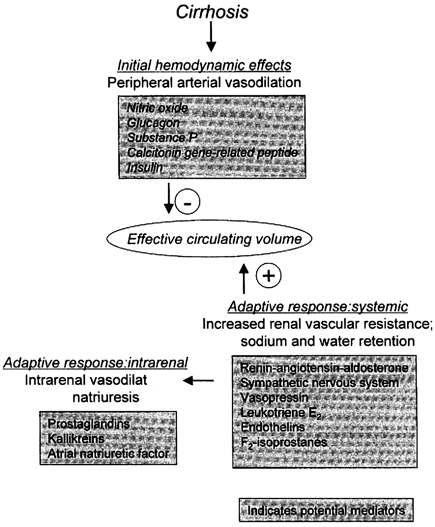
Sodium resorption increase caused by vasoconstriction leads to the decrease of sodium transport to distal tubulus, where aldosterone and atrial natriuretic peptid work. Specific factors, which take part in initial peripheral artery vasodilatation, remain unclear. The levels of glucagon in plasma; substance P (4) and calcitonin in plasma are higher. Calcitonin, genetically connected peptide, is a potent vasodilatator, and its level in circulation is increased in patients with alcoholic cirrhosis and ascites, but not in the control group of patients with cirrhosis but without ascites. Regardless of etiology, vasodilatation of peripheral arteries and insufficient arteries filling initiates a response aimed at recovering artery pressure (3). The activation of sympathic nervous system, renin-angiotensin-aldosterone system, (7) vasopressin and the other factors together lead to the increase of renal vascular resistance (3) and renal sodium and water retention (8). It seems that the explanation for the damaged possibility of water excretion is nonosmotic stimulation due to arginine vasopressin leaving, which characterizes advanced cirrhosis. There is a higher level of vasoconstrictor endotelin-1 in plasma in more conditions characterized by sodium retention (9). In healthy individuals the infusion of small doses of endothelin 1 decreases sodium excretion for 36%. These effects can be partly blocked by calcium antagonist nifedipine. The level of endothelin 1 and endothelin 3 in hepatic arteries and veins is increased in patients with liver cirrhosis (9). The values are higher when ascites is also present. The level of endothelin 1 and endothelin 3 in serum correlates with significantly higher values of serum creatinine (9) and negatively correlates with central and artery blood volume, diastolic blood pressure and sodium values in serum (9).
DIAGNOSIS
Diagnostic criteria for HRS
HRS should thought to be present in all patients with acute and chronic liver illness and portal hypertension when there is a growth of serum creatinine over 1.5 mg/dL. A HRS appears in the presence of relatively advanced liver disease and the risk of a HRS is higher in patients with hyponatremia, high activity of renin in plasma and decreased liver. Neither the etiology of the illness nor Child-Turcotte-Pugh has a significant importance in the anticipating of this disease (10). The International Ascites Club summarized criteria for the diagnosis of HRS are shown in the diagram 2 (1). The criteria were grouped into: major criteria necessary for setting the diagnosis and minor criteria, which represents the support in setting the diagnosis. Major criteria include: 1) the presence of acute or chronic liver disease with the advance of the hepatic failure and portal hypertension; 2) a low glomerular level filtration rate, which is marked by the amount of creatinine in the serum higher than 1.5mg/dL or clearance creatinine lower than 40ml/min; 3) the absence of the use of nephro-toxic medicines, shock, infection, a recent loss of liquids or some other cause of nephrotoxicality; 4) lack of significant improvement of kidney function following diuretics withdrawal and volume expansion with 1.5 L of isotonic saline; and 5) no evidence of a significant proteinuria, renal obstruction or parenchymal disease of kidneys (1). Minor criteria are mainly based on the functional evidence of low glomerular filtration and avid sodium retention including: 1) urine volume less than 500 ml/24h; 2) sodium concentration in the urine less than 10 mEq/L; 3) urine osmolality higher than plasma osmolality; 4) absence of significant hematuria; and 5) sodium concentration in the serum less than 130 mEq/L. Although they are not necessary for setting the diagnosis; the absence of these criteria can raise doubts about the set diagnosis (18). In patients with a susceptive HRS other possible causes for kidney insufficiency should be looked for. It is also necessary to do an ultrasonography examination in order to exclude parenchymatous disease of kidneys (18). One of significant problems in the early diagnosing of a HRS is that there are significant restrictions in the amount of serum creatinine as a marker of a renal function. The assessment of glomerular filtration on the basis of the amount of serum creatinine and clearance creatinine overestimates the real value of glomerular filtration in the presence of cirrhosis. For instance, comparing the clearance of inulin in 56 cirrhosis patients, the sensitivity of serum creatinine (18%) and clearance of creatinine (74%) in the kidney, it was obvious that insufficiency detection weren't significant; these tests overestimate the real value of glomerular filtration in about 50% of persons with a reduced clearance of inulin. This overestimation appears as the result of the increased tubular secretion of creatinine (11). The values of serum creatinine and clearance of creatinine don't seem to be an adequate index of the kidney function even in patients with well compensated cirrhosis. In 68 patients with uncomplicated cirrhosis who are not in azotemia the evidence of decreased renal function was found in almost two thirds of them, including 21 patients with clearance of creatinine of 50-80 ml/min and 25 patients with clearance of creatinine of less than 50 ml/min. The average monitoring of 180 days indicated that the rate of mortality was 24% in patients with clearance of creatinine of 50-80 ml/min and 36 patients with clearance less than 50 ml/min comparing to 9% in patients with normal kidney function. The use of Doppler ultrasonography aimed at establishing the index of renal vascular resistance is suggested as an alternative method for early diagnosing of a HRS. In cirrhosis patients with the kidney insufficiency the index of resistance correlates with the level of glomerular filtration, artery pressure and renin activities in plasma (12) and is both sensitive and specific for diagnosing kidney weakness in 71 - 80% (12). However, this method is not applied as routine in clinical procedures.
TREATMENT
The treatment of a HRS mainly begins with diuretics use within the treatment of portal hypertension and ascites. This treatment also includes rest, less sodium input, intensive use of diuretics, especially spironalacton (400 mg/24h); some patients also need the additional treatment with furosemide (160 mg/24h) (1). These measures lead to the control of ascites genesis in 85% of patients, but there is a risk of pre-renal azotemia, hypokaliemia and encephalopathy. Patients, who do not respond to this treatment after a week; probably have refractory ascites, which often leads to a HRS. The growth of serum creatinine level to the value of 1.5 mg/dL under these circumstances needs the intensive treatment. Initially, all potentially nephrotoxic agents including nonsteroidal antiinflammatory drugs, aminoglycosides, and other antibiotics must be excluded. Pre-renal azotemia can disguise clinical and laboratorial tests of a HRS, including oliguria, low sodium concentration in urine and high urine osmolality. Since there is a high risk of pre-renal azotemia incidence caused by digestive bleeding or some other liquid loss in patients with liver cirrhosis, it is necessary to do the compensation of volume in the most of them (18). The most significant restriction of this treatment is the resistance to diuretic treatment, which probably appears due to the loss of mineralocorticoids (13). Evacuation of large quantity of ascites may substantially increase the risk of pressure fall in the portal system as well as of renin and aldosterone level decrease in plasma, which can lead to the growth of the level of the nitrogen products in serum due to effects on intravascular volume. There is restricted data about extracorporal albuminic dialysis and treatment with law dosis of dopamine, combination of norepinephrine and dopamine (13), terlipresin (14), and analog of vasopressin omnipresent. Although there are studies concerning the application of these medicines, they are still not widely used in clinical procedures. Several cases have shown a possible, treatment role of transjugular portosystemic shunt (TIPS) in HRS (15). In the series of patients with refractory ascites, TIPS increases sodium excretion through urine and improves serum creatinine level as well as glomerular filtration level. These consequences are related to the decrease of aldosterone level in plasma and renin activities. Clinical improvement of ascites has been found in 74% of treated patients and the average dose of diuretics treatment has been decreased for 50%. Most patients have not had a recurrent HRS. Therefore, TIPS can be an acceptable solution for patients who are resistant to diuretics treatment although its role in HRS is still to be established. Kidney insufficiency incidence in patients with cirrhosis has a very bad prognosis. Renin activity in plasma, anti-diuretic hormone concentration and sodium concentration in serum have a certain value as prognosing factors (12). The traditional attitude is that dialysis is not effective at all in patients with a HRS, except when it is used to cover the period of the liver transplantation (17). Patients who already have a liver disease and acute kidney insufficiency need dialysis though the rate of mortality is especially increased when there is thrombocytopenia (the number of palatals is bigger than 100.000 mm3), hepatitic encephalopathy or prolonged prothrombin time. When there are no complications like these, the survival period of one year is 38% (17). The kidney insufficiency itself in the absence of counter-indications does not have to exclude the dialysis though there is a significant risk. An orthotopic liver transplantation remains as the last possible treatment for a HRS. When it is successful, a complete kidney insufficiency recovery can be expected. Transplantation postponement until the kidney insufficiency appearance, significantly increases the risk. A recipient with a HRS has a significantly shorter a five-year period of survival as compared to the ones without a HRS. These patients should be hospitalized for as long as possible; they should also be placed in the intensive care for as long period as possible with frequent dialyses. An early transplantation represents the best option for these patients.
REFERENCES
1. Arroyo V, Gines P, Gerbes AL, et al. Definition and diagnostic criteria of refractory ascites and HRS in cirrhosis. Hepatology 1996; 23:164-176.
2. Gines P. Diagnosis and treatment of HRS. Baillieres Best Pract Res Clin Gastroenterol 2000; 14: 945-957.
3. Badalamenti S, Graziani G, Salerno F, Ponticelli C. HRS. New perspectives in pathogenesis and treatment. Arch Intern Med 1993; 153:1957-1967.
4. Lang F, Gerok W, Haussinger D. New clues to the pathophysiology of hepatorenal failure. Clin Invest Med1993; 71:93-97.
5. Epstein M. HRS. Emerging perspectives of pathophysiology and therapy. J Am Soc Nephro1994; l 4:1735-1753.
6. Gupta S, Morgan TR, Gordan GS. Calcitonin gene-related peptide in HRS. A possible mediator of peripheral vasodilatation? J Clin Gastroenterol 1992; 14: 122-126.
7. Bernardi M, Trevisani F, Gasbarrini A, Gasbarrini G. Hepatorenal disorders: Role of the renin-angiotensin-aldosterone system. Semin Liver Dis 1994; 14:23-34.
8. Gines P, Arroyo V, Rodes J. Ascites and HRS: Pathogenesis and treatment strategies. Adv Intern Med 1998; 43:99-142.
9. Moller S, Gulberg V, Henriksen JH, Gerbes AL. Endothelin-1 and endothelin-3 in cirrhosis: Relations to systemic and splanchnic hemodynamics. J Hepatol 1995; 23:135-144.
10. Platt JF, Elis JH, Rubin JM, et al. Renal duplex Doppler ultrasonography: A noninvasive predictor of kidney dysfunction and hepatorenal failure in liver disease. Hepatology 1994; 20:362-369.
11. Caregaro L, Menon F, Angeli P, et al. Limitations of serum creatinine level and creatinine clearance as filtration markers in cirrhosis. Arch Intern Med 1994; 154:201-205.
12. Maroto A, Gines A, Salo J, et al. Diagnosis of functional kidney failure or cirrhosis with Doppler sonography: Prognostic value of resistive index. Hepatology 1994; 20: 839-844.
13. Durkin RJ, Winter SM. Reversal of HRS with the combination of norepinephrine and dopamine. Crit Care Med 1995; 23:202-204.
14. Hadengue A, Gadano A, Moreau R, et al. Beneficial effects of the 2-day administration of terlipressin in patients with cirrhosis and HRS. J Hepatol 1998; 29:565-570.
15. Sturgis TM. HRS: Resolution after transjugular intrahepatic portosystemic shunt. J Clin Gastroenterol 1995; 20:241-243.
16. Yersin B, Burnier M, Magnenat P. Improvement of renal failure with repeated head-out water immersions in patients with HRS associated with alcoholic hepatitis. Am J Nephrol 1995; 15:260-265.
17. Keller F, Heinze H, Jochimsen F, et al. Risk factors and outcome of 107 patients with decompensated liver disease and acute renal failure: The role of hemodialysis. Ren Fail 1995; 17:135-146.
18. Sleisenger M, Friedman L, Feldman M. (eds). Hepatorenal syndrome. In: Gastrointestinal and liver disease. 7th edition Saunders 2002; 562-564.