|
|
Original article
BIOPHYSICAL PROCESSES AND ANGULAR TRACTION EFFECTS DURING FETAL VACUUM
EXTRACTION
Folić Miroslav1, Čitaković Ivanela1 , Jeremić Branislav2
1 Obstetrics and Gynecology Clinic, Clinical Center Kragujevac
2
Terotechnology Institute, Mechanical Engineering Faculty Kragujevac
INTRODUCTION
Fetal vacuum extractor is used in 0.9-5.2% of births (1,2,3). The rationale of these differences in frequency of FVE use are conditioned by the attitudes of obstetric clinics towards the completion of expulsion delivery phase during fetal head stoppage over the pelvic exit and the consequences which the chosen delivery termination method may lead to (4,5). Investigation of these situations are the part of most up-to-date multidisciplinary investigation tendencies and they connote extensive use of medical engineering. This includes the application of complex knowledge from the fields of medicine, theory of the model formation process, biophysics, electronics, mechanics, measurement theory etc. This original scientific paper is the result of complementary investigations of the Clinic of Gynecology and Obstetrics and the Institute of Terotechnology, Machine Engineering Faculty in Kragujevac, for the first time conducted in Serbia and Montenegro, with quantification of the processes of biophysical modeling during vacuum fetal extraction and simultaneous determination of the angular traction effects.
AIM OF THE WORK
The aim of the work is objectification of the biophysical processes occurring on the fetal head during vacuum extraction, investigation of the potential consequences of negative pressure applied on the angular surface of fetal head, determination of traction forces as well as determination of the conditions for evaluation of posttraumatic decompression effects.
RESULTS OF THE INVESTIGATION
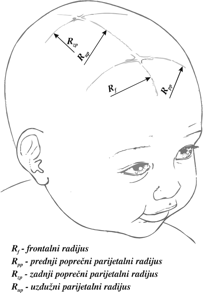
Modeling of the biophysical processes during fetal vacuum extraction (FVE) connotes work in several phases. The first phase is an exact definition of anatomical characteristics of fetal scalp. Measurements were made on a representative sample - the characteristic radius values were shown in the annex figure 1 and the numerical values in table 1.
|
|
||||||||||||||||||||||||||||||||||||||||||||||||||||||||||||||||||||||||||||||||||||||||||||||||||||||||||||||||||
| Table 1. Numerical values of the fetal head radius | |||||||||||||||||||||||||||||||||||||||||||||||||||||||||||||||||||||||||||||||||||||||||||||||||||||||||||||||||||
Fetal scalp, according to these measurements, can be modeled as a geometric spherical form, with diameter of 100 mm; based on that, an investigational rubber model was made, with the attached dynamometers to measure traction force and traction momentum (demonstrated in figure 2).
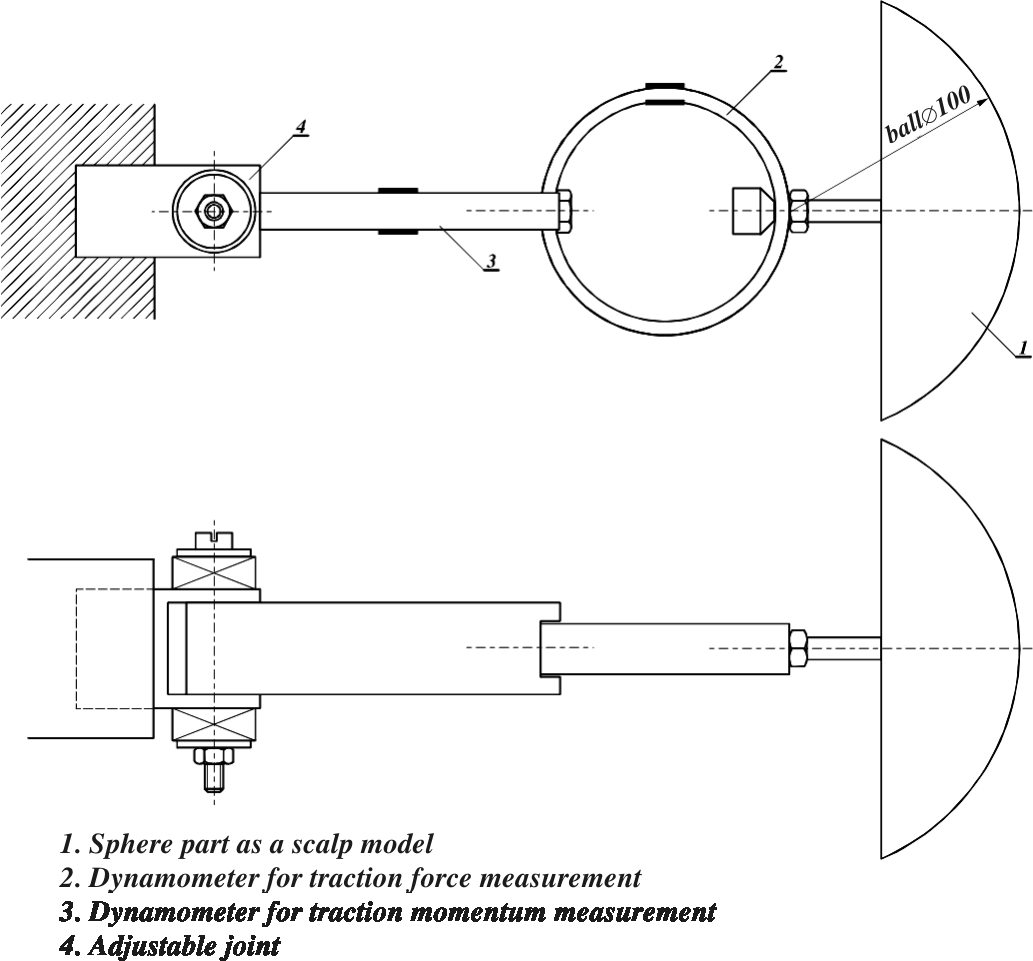
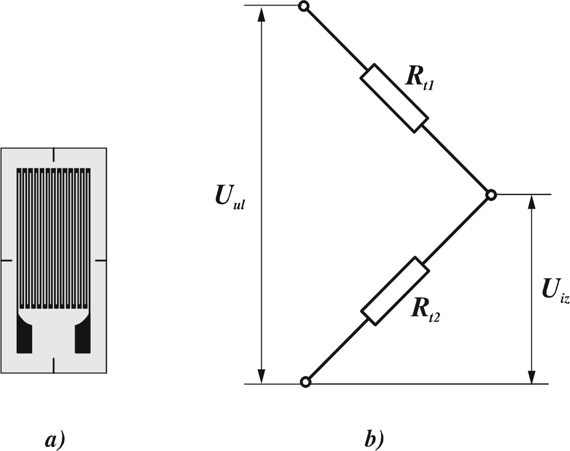
Figure 2. Model of the fetal scalp with dynamometers
Dynamometers to measure the traction force and traction
momentum in FVE were made of measurement bands based on the tensoresistive
effect, the phenomenon that if a metal conductor is deformed its resistance is
altered too. Using the Hook’s law concerning the association of charge and
deformity (s
= E × e)
and charge and force, s
= F / A (where A stands for the surface upon which the force is applied), a
relationship may be obtained between the force acting upon the conductor and
alte ration of its resistance. Measurement stripes are specially devised
conductors between two leafs of plastic attached to the object the deformities
of which are to be measured (figure 3). Since the measurements were performed at
the room temperature, there was no need to eliminate the temperature effect on
the resistance alteration - measurement stripes were tied in the Witson ring
half-bridge.
|
|
|
|
Figure 3.
Measurement stripes (a); |
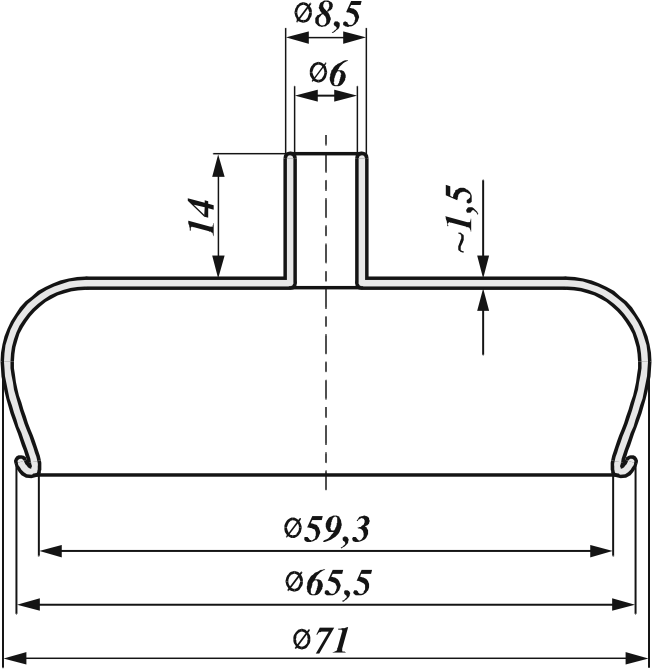
Figure 4. Calotte of the vacuum device |
On deformation and alteration of resistance of measurement stripes, the
output charge of the bridge Uiz is altered. These changes represent the
measurement of deformity. Dynamometers are devised and manufactured depending on
the intensity and type of force to be measured. Before measurements, it is
necessary to burden them with known forces and determine functional dependence
of the output charge.
Calculation of the UM charge obtained with the dynamometer in the position 3,
figure 2, into the traction momentum is performed according to:
Mt = KM × UM
where KM (Nm/V) is the proportion factor depending on the applied measurement stripes, transversal surface of the dynamometer and arms of the force applied. The charge obtained with dynamometer marked with position 2, figure 2, is expressed as:
U2 = k1 MT + k2 × Fa
in which k1 and k2 are influence coefficients. Finally, determination of the traction force Fa could be obtained with the expression:
![]()
An experienced obstetrician and numerous applications of fetal vacuum
extractor defined empirically the resistance values with the adjustable joint
(figure 2, position 4). This adjustable joint is a tribomechanic system with the
elements among which a versatile traction force is acting, depending on the
magnitude of contact pressure on the elastic joint slugs. These slugs are placed
in order to enable a continual regulation of the resistance simulated during the
FVE process modeling.
The magnitude of the contact pressure may be changed by the change of force
of tightening the screw which keeps the slugs within the joint; it could be
quantified as follows:
F = c x,
in which c is stiffness of the elastic slugs and x deformity of the slugs. The force of tightening the screw makes a contact pressure onto the elastic slugs according to the formula:
![]()
where A is the contact surface between the slugs and the joint carrier.
In the first approach the tightening force F may become equal as the normal force Fn acting in the joint onto the contact surface between the elastic slugs and the constrained joint carrier, so:
F = FN
Between the contact surfaces of this tribomechanic system a traction force is acting
Fµ=µ FN ,
in which µ is the traction coefficient value, completely equal.
Therefore, by the contact pressure change (ie. force within the joint) altered is also the traction force an obstetrician should overcome during the traction process. Simulation of the traction momentum is performed through the MT:
MT = Fr l,
where Fr is the radial FVE force component.
The MT momentum fetal head is brought into the flexion position defined by
the negative á angle values, ie. the deflexion defined by positive á values.
FVE device creates a vacuum, transmitted through a rubber hose to the fetal
scalp. Vacuum device calotte transmits the traction force to the fetal scalp
made by the physician. Maximal transmittable force is the function of the
calotte surface A and the sub-pressure Dpv achieved by the vacuum device:
F=D pv A
The level of vacuum in FVE conventionally does not exceed 0.9 bar. Therefore
there are several sizes of vacuum device calotte which enable that the maximal
force could be different. In the figure 4, some basic calotte dimensions were
shown used in experimental studies, and in the figure 5 models of the reduced
pressure vacuum chamber for flat, spherical and supposed deformed contours of
the scalp contact surface.
Maximal force which may be generated with the given pressure level could be
calculated accor- ding to the expression F=DpvA and relates to the vacuum
chamber with flat contact surface, while in all other supposed deformities
maximal force could be calculated as follows:
![]()
with A as the surface defined by the diameter D (figure 5).
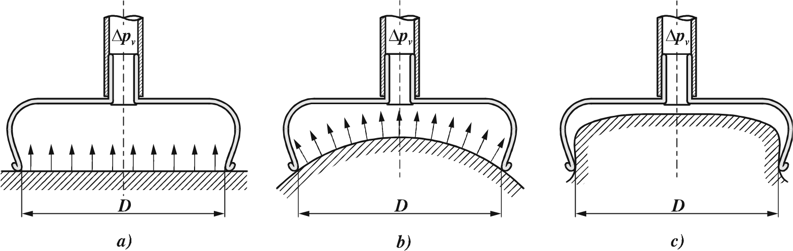
Figure 5. Formation of the vacuum chamber: contact surface flat (a), spherical (b) and deformed (c)
It should be emphasized that the fetal scalp modeling was performed with the part of a rubber sphere, the deformities of which could be disregarded in view of the actual scalp deformities. In real situations, a part of the fetal scalp is retracted into the FVE vacuum device calotte (figure 5c). On retraction, there is a possibility that for a short period a holding force is effectuated larger than the theoretically calculated force; to the vacuum exerted holding force a friction force (between the retracted fetal scalp and vacuum calotte) should be added.
In the table 2 some maximal, theoretical values of the vacuum holding force are given, calculated according to the above mentioned expression, as well as the maximal measured values of the holding force (with different vacuum values).
Table 2. Maximal vacuum holding force values
|
Sub-pressure Dpv |
Maximal theoreti-cal holding force |
Maximal mea-sured holding force |
|
bar |
daN |
daN |
|
0,70 |
19,33 |
16,71 |
|
0,75 |
20,71 |
17,49 |
|
0,80 |
22,09 |
18,41 |
|
0,85 |
23,47 |
19,29 |
|
0,90 |
24,86 |
20,60 |
|
0,95 |
26,24 |
22,47 |
In the figure 6 the forces acting upon the rubber model through the calotte are presented.
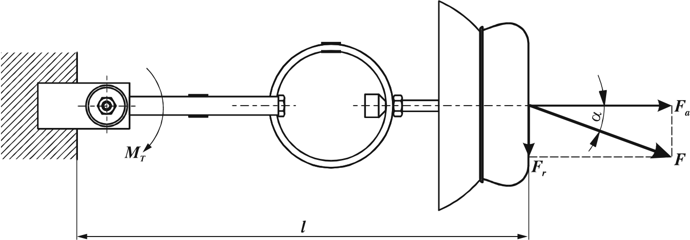
Figure 6. Forces acting through the FVE calotte onto the scalp model
The F force with which an obstetrician retracts the calotte acts at an angle á to the axis corresponding to the calotte axis. That force can be divided into two components: Fa, traction force (acting along the calotte axis – axial force) and Fr component (acting perpendicularly to the calotte axis – transversal force). The transversal force creates a separate traction momentum calculated after the expression:
M = MT = Fr × l
A measurement chain for traction force and momentum monitoring is shown in figure 7.
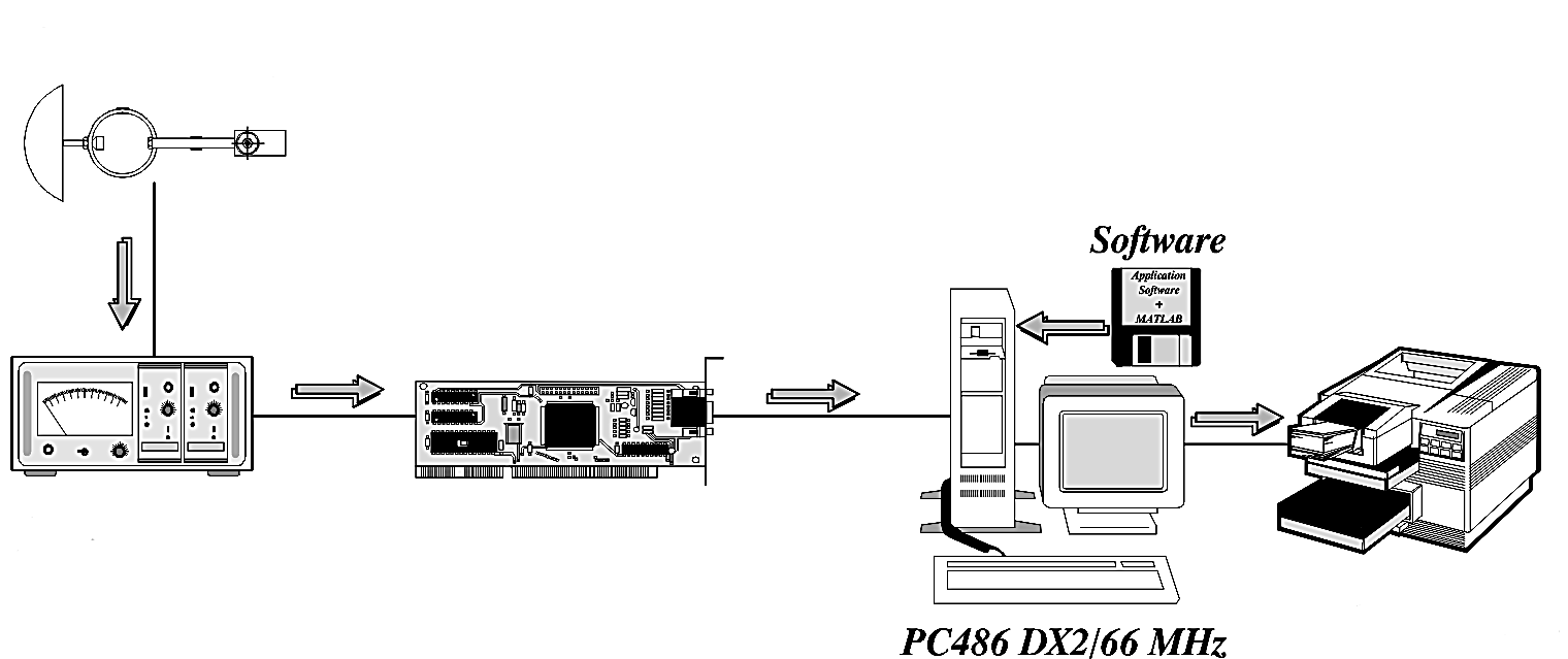
Figure 7. Measurement chain for traction force and momentum monitoring
The signal from the measurement dynamometer stripes (conveying the
information on the traction force and momentum) is transmitted to the
two-channel bridge amplifier, where the signal is augmented to the level
necessary for A/D converter function. A/D converter is built in a personal
computer; its role is to convert continual electric signal into a sequence of
numbers (discrete values). A signal in the form of numerical sequence is
appropriate for further numerical processing. Software for A/D converter control
is the LABTECH NOTEBOOK model, while for signal processing and presentation
Matlab for Windows was used.
Figure 8. Delivery process according to the angular traction phases
In figure 8, a delivery process is demonstrated by the angular traction phases.
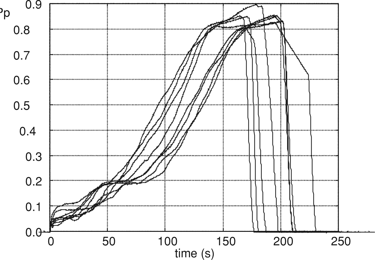
Check-ups of the subjective status of an obstetrician which performed several hundred FVE applications was performed through the process repetition on the model, with the obstetrician who could not monitor the values of forces and FVE timing. The result of this study is a family of curves through which formation of the traction vacuum is monitored (figure 9), demonstrating very good reproducibility. Based on clustering, a representative curve was devised (figure 10).
 |
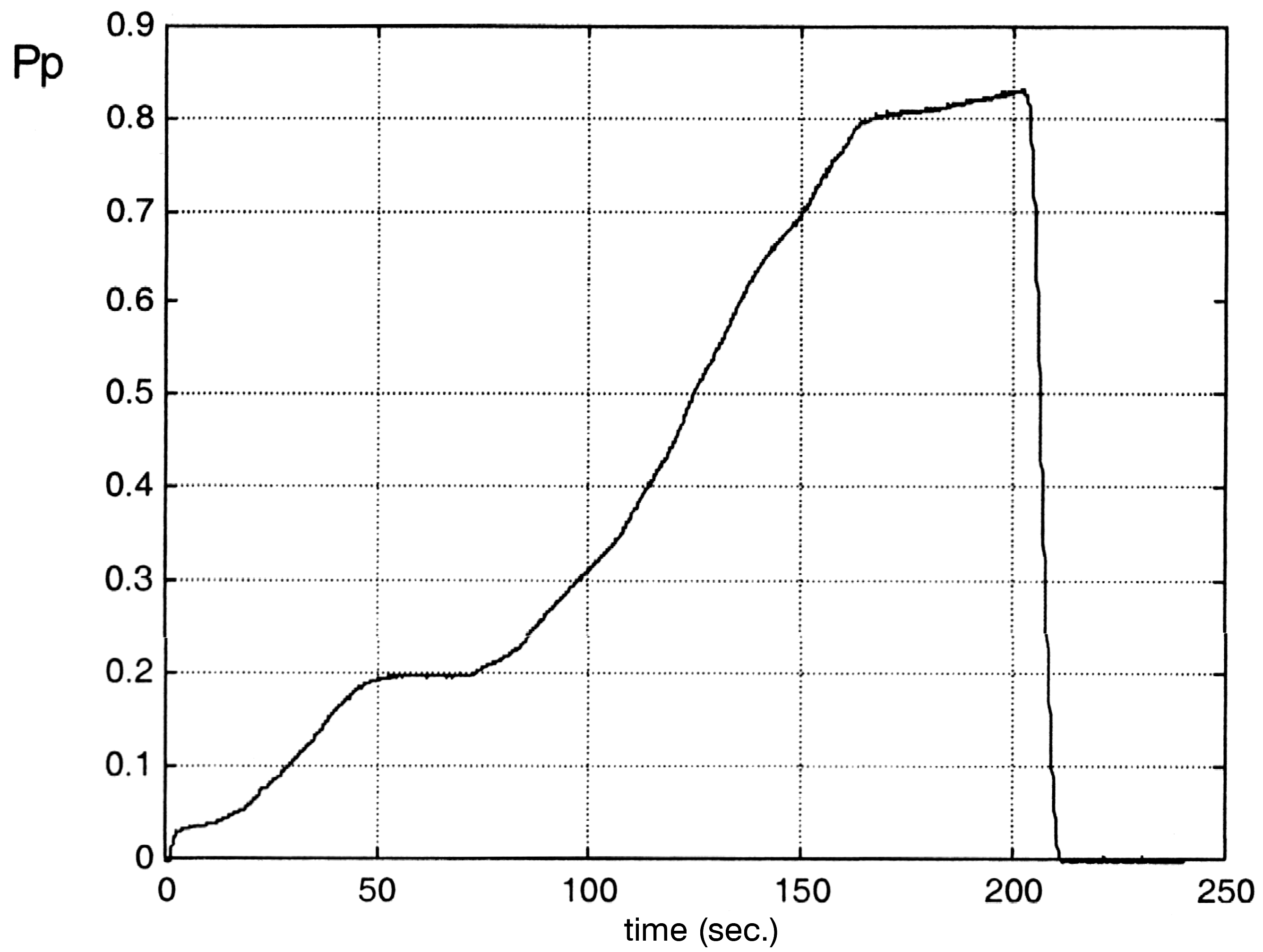 |
| Figure 9. Family of the curves of traction vacuum formation | Figure 10. Representative curve of the traction vacuum formation; initial vacuum formation phase(up to 50 s), revision of the angular application (up to 100 s), forced traction vacuum formation(up to 170 s) and the phase of angular traction revision and extraction (up to 220 s) |
CONCLUSION
Investigation of the biophysical processes occurring on the fetal head during FVE and the effects of angular tractions confirm the following opinions:
1. The force created (maximal theoretical is 26.24 daN, and maximal measured
22.47 daN) must not be used to resolve the problems of disproportion between the
fetal head and osseous pelvic delivery route.
2. The success of FVE placement is directly proportional (which follows from
the first conclusion) to the accurately established relationships of these two
parameters.
3. FVE assisted delivery should be performed only by an experienced
obstetrician (several years experience; multiple FVE applications), with the
appropriate knowledge of biophysical processes and angular traction effects, and
with
4. Reliable neonatology and anesthesiology support team, well trained,
equipped (laboratory and instruments/machines) and always ready to render top
quality support to the mother and her newborn child.
Future studies of the biophysical processes in FVE and consequences of angular traction should clearly define deformity processes on the fetal scalp and quantify the phenomena in the intra-cranium, especially elevation of the intracranial pressure and hemorrhages in the brain structures (above all in the ventricles), as a leading partal risk in instrumentally assisted delivery with FVE.
LITERATURA
1. Grossman, RA. An easily made obstetrical vacuum extractor. Am Med J Obstet
Gynecol 1999; 22(4):113-20.
2. Johanson RB, Rice C, Doyle M, et al. A randomized prospective study
comparing the new vacuum extractor policy with forceps delivery. Br J Obstet
Gynecol, 2001; 100 (6):524-30.
3. Lucas, M J. The role of vacuum extractor in modern obstetrics. Clin Obstet
Gynecol, 2001; 37 (4): 794-805.
4. Muise K L, Duchon M A, Brown R H. The effect of artificial caput on
performance of vacuum extractors. Obstet Gynecol. 2002. 88 (6): 122-34.
5. Vacca A. Effect of angular traction on the performance of modern vacuum
extractors. Am J Obstet Gynecol. 2001; (9), 500-09.