|
|
Original article
CLINICAL AND RADIOLOGICAL FORMS OF SARCOIDOSIS OF LUNG
Tatjana Pejčić1, Ivana Stanković1, Milan Rančić 1, Ivanka Đorđević1, Ljiljana Isaković2, Lidija Ristić1
1 Clinic for lung disaese and tuberculosis, Clinical center Niš,
2 Special hospital for
rehabilitation of lung diseases, Sokobanja
INTRODUCTION
Sarcoidosis is a systemic granulomatous disorder of unknown etiology,
characterized by bilateral hilar adenopathy (BHA), lung parenchyma
infiltrations, skin and eyes changes or other organs involvement. Histological
characteristic of sarcoidosis is the presence of non- caseating granulomas,
which can resorb or evolve into fibrosis. Immunological tests show a depression
of delayed hypersensitivity reaction, amplified activity of Th1 immune response
on affected organs, hyperreactivity of B lymphocytes and elevated values of
circulated immune complexes (1).
This disease is well-known around the world, yet, with quite different
prevalence and incidence ranging. In Sweden the prevalence ranging is 64/100000,
in Norway 26,7/100000 and in USA 11/100000. There is no precise data about the
frequency of sarcoidosis in Serbia and Montenegro, but the areas of Sandzak and
West Serbia (2,3) are considered to be predilactive. As early as 1905 M. Boeck
described sarcoidosis as "a bacillary infections disease ", but until now its
etiology hasn't been explained. The possible etiological causes are induced
unorgans agents (cirkonium, aluminium, talc), viruses (herpes virus 8,
rethrovirus, Epstein- Barr virus), mycobacteria (M. tuberculosis, M.
paratuberculosis, other mycobacteria) and other bacterias considered to be
capable of producing granuloma. Until now, there have been no infective agents
isolated from affected tissue; so, this hypothesis is questionable. Sarcoidosis
frequently affects younger women between 20-30 years of age. A family connection
was observed as far as illness is concerned, with more frequent relation between
brother and sister as well as mother and children. This confirms the theses
about the presence of genetics in sarcoidosis appearance. The connection between
sarcoidosis and HLA-1, HLA-B8 and HLA-DR4 has been confirmed. A good clinical
outcome of disease is associated with HLA-D3, whereas only pulmonary sarcoidosis
is associated with HLA-B27 (4,5).
Depending on the period of symptoms appearing, sarcoidosis is clinically
classified as acute and chronic form. Acute forms are characterised by symptoms
over two year period, and chronic one by symptoms lasting more than two years.
General symptoms of disease are frequently expressed by fever, weakness, loss of
weight, while specific symptoms of disease are related to organ involvement. In
more than a half of the patients respiratory tract is affected; so, the most
dominant symptoms are dry cough and chest pain. The most common acute
manifestation of sarcoidosis is Lofgren's syndrome, characterized by fever,
weakness arthritis, erythema nodosum and BHA on chest x - ray. Sarcoidosis is
considered as a disease with good prognosis; 90% of the patients show
spontaneous remission of all symptoms in a two year period, and also, there are
cases with permanent BHA. The most predominant extrapulmonary symptoms of acute
sarcoidosis are eye disorders (conjuctivitis, photophobia and sight disturbance)
and skin disorders. Sarcoidosis of cardiac and abdominal organs and changes of
central nervous system are substantialy rare. Pheripherial lymphoadenopathia can
be present in 60-75%. Erythema nodosum and acute inflammation manifestation
(fever and polyarthritis), are indicators of good prognosis (2,6).
Chronic course of sarcoidosis is present in patients with symptoms lasting
more than two years. On the basis of literature data, chronic sarcoidosis
appears in about 10-30 % of the patients. Spontaneous remission in this group
occurs in two thirds of the patients, and thus, chronic course of disease
persists in 10-30% of the patients.
Severe extrapulmonary localisation of sarcoidosis (involvement of myocard,
CNS, liver, spleen) is present in 4-7% of the patients in the onset of disease
and its atypical beginning are the first signs of bad prognosis and chronic
course of disease (7).
Symptoms of chronic sarcoidosis depend on organs involvement and functional
symptoms. On the basis of radiographical involvement of pulmonary sarcoidosis
(Wurm - Reindell - Helmeyer) the first stage is characterised by bilateral hilar
lymphadenopathy, the second one by BHA as well as lung parenchyma infiltrations,
whereas the third one is characterised only by parenchymal infiltrations with or
without interstitial fibrosis, and finally the fourth refers to radiological
stage including fibrous lines spreading up from the hilus. Relaps of the disease
is defined as repeated appearance of symptoms and signs of disease after the end
of previous treatment or as reappearance of disease besides treatment of
morbostatic doses of glucocorticoides (5-10mg) (7,8).
THE AIM
The aim of our paper was to show the most frequent clinical and radiological forms of pulmonary sarcoidosis in patients treated at the Clinic for lung disease and tuberculosis in Knez Selo. Particular attention was paid to pulmonary functional tests, with reference to mutual correlation of some lung functional parameters.
METHODS
We retrospectively analyzed medical and/or hospital records of 45 patients
reported to Clinic for lung disease with a suspicion of having pulmonary
sarcoidosis. Patients were from 30 to 62 years old, with some higher number of
women (25) in relationship to men (20). We tested the onset of disease, duration
of symptoms, the most frequent pulmonary, extrapulmonary as well as general
symptoms of disease.
We analyzed chest radiography (rtg) and classified radio-graphical staging of
disease.
All patients underwent pulmonary functional tests which presupposed
spirometry and flow-volume curve as well as determining diffusing capacity of
lungs for carbon monoxide (DLco) (Pneumo screen/ Diffusion firm Jaeger).
On the basis of values of vital capacity (VC) and forced expiratory volume in
the first second (FEV1) as well as middle expiratory flow at 50 percent of VC
(FEF50) and FEF75, disturbance in ventilatory function were determined. We
analyzed the correlation between lung functional parameters (FEV1, VC) and
values of DLco.
From statistical parameters middle value (x), standard deviation (SD) and
coefficient of correlation (r) were used.
THE RESULTS
The examination covered 45 patients, between 30 and 62 years of age (x
41,2±10,6), 25 women (55,5%) and 20 men (44,5%). The diagnosis of sarcoidosis in
10 patients confirmed the seizure of organs after biopsy and histological
verification of changes in lymph nodes or lung parenchyma, and in 35 patients as
probable criteria (without histological confirmation, but with symptoms and
signs of sarcoidosis and presence of lymphocytes alveolitis in bronchoalveolar
larvate (BAL) and lowered values DLco and other factors of sickness activities)
(9).
In 30 patients (66,6%) the beginning of the disease was acute with Löfgren's
syndrome (higher temperature, erythema nodosum and BHA). One patient had
enlarged parotids glands, front uveitis, higher temperature and paralysis nerves
facials (Heefort- Waldenstrom syndrome). These patients had been having those
symptoms less than two years.
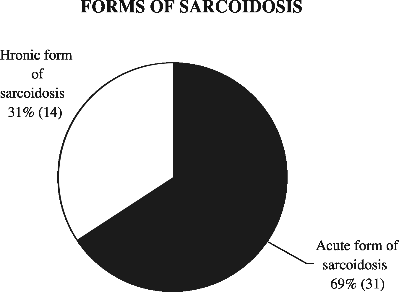
14 patients (31,2%) had symptoms more than 2 years and we marked them as
those with chronic disease (figure 1).
|
|
|
|
Figure 1. Forms of sarcoidosis |
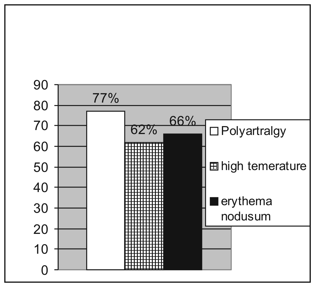
Figure 2. Non lung symptoms in sarcoidosis |
Besides lung symptoms patients also had: polyarthralgia (35/ 77%), mostly in
wrists (30/ 66,6%), and palm wrists of both hands (10/22,2%), higher temperature
(28/62%), sub febrile type, erythema nodosum 30 patients (66,6%) (figure 2). All
patients were complaining of weariness and tiredness.
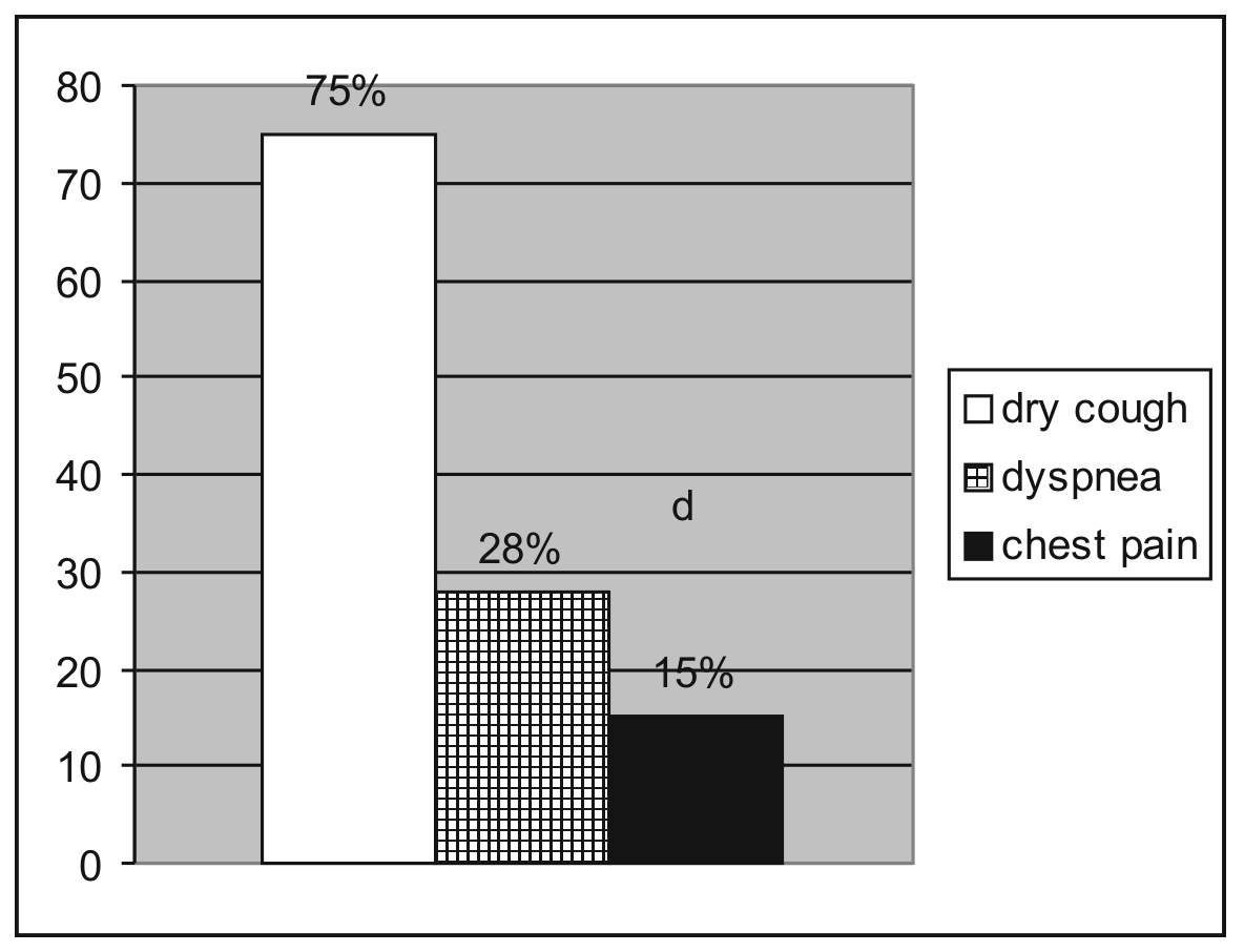
As far as extrapulmonary symptoms are concerned, patients mostly complained
of dry cough (34/75,5%), dyspnea (13/ 28,8%) and chest pain (7/ 15,5%) (figure
3). Patients complained mostly of chest pain lasting more than 1 year in the
case of 5 patients (11,1%), and the feeling of weariness (25/ 55,5%) as one of
the general symptoms.
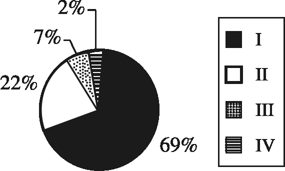
31 patients (68,8%) had radiological report of the I stadium of lung
sarcoidosis. One patient had only one sided hilar adenopathy, and other had both
sided hilar and mediastinal, 10 patients (22,2%) had II radiological stadium, 3
patients (6,66%) had III and 1 patient (2,2%) had IV radiological stadium of
sarcoidosis (figure 4).
|
|
|
|
Figure 3. Lung symptoms of patients in sarcoidosis |
Figure 4. Radiological stadium of sarcoidosis |
30 patients (66,6%) had preserved lung ventilation (28 patients with acute stadium and 2 patients in the second radiological stadium). Changes in small lung airways (FEF50 and FEF75 < 60% of expected values) had third patients (6,6%) (two with acute sarcoidosis and one with chronic sarcoidosis in II radiological stadium). Five patients had obstructive disorder of ventilation (11,1%) with FEV1 from 52% to 74% (x 61,11± 11,2) (one of the patients was with I rtg stadium and four with II rtg stadium). Seven patients had restrictive disorder of ventilation (15,5%) (one in IV rtg stadium, three in III rtg stadium and three in II rtg stadium of illness). The values of VC were from 51% to 76% (x 63,1 %± 8) (figure 5).

Figure 5. Lung function test in sarcoidosis
DLco was lowered in 30 patients and values were from 46,9% to 70% (x 62,8%± 7,3 ). In one third of the patients (10 patients) with preserved lung ventilation, values of DLco were lowered below 70%. No significant statistical correlation of parameters VC and FEV1 with DLco (r =0,34, p> 0,05, and r= 0,37, p> 0,05) were found. In one patient in III rtg stadium of illness and chronic sarcoidosis, sarcoidosis of myocardia and pancreas was proved.
DISCUSSION
Immunopathogenesis of sarcoidosis includes initial agents as a possible
trigger for the beginning of disease. CD4+ T lymphocytes and mononuclear
phagocytes have the leading role in that. The sarcoid granuloma is considered to
be the consequence of induced immune response to unknown agent which persists on
active affected sites, probably due to low solubility or demolition.The first
pathological manifestation of disease is accumulation of immunocompetent cells
(mostly CD4-lymphocytes and macrophages) followed by producing lymphocytes
alveolitis. The next step is the formation of granulomas, which originates from
high- differentiated mononuclear phagocytes (epitheloid and multinucleated
gigant cells) and lymphocytes. The giant cells are formed by IL-2, IFN g and IL
1ß. In the centre of sarcoid granuloma CD4+ cells are dominated, and on the
periphery there are scatterd CD8+, CD4+, B cells, fibroblastes, mastocytes and
other cells. The evidences pointed out that sarcoidosis is a consequence of
defective cellular immune response, triggering by specific antigen related to
genetic disorder. In the early phase of disaese, at the site of sarcoid
lessions, there is marked enhancement of number of Th1 lymphocytes, which
cytokines increased the growth of granuloma by inhibition of fibrosis. Depending
on organism tendency, getting into Th2-lymphocites response leading to
progression of sarcoidosis and appearing of fibrosis. By cytokines realising
throught Th2 lymphocytes fibroblaste hyperplasio originated with amplified
formation and deposition of extracellulare matrix components in the surrounding
granulomas inflamation (6,4,1).
We showed 45 patients with sarcoidosis, among whom the most dominant were
those with acute form of illness in I rtg stadium (31/ 68,8%). According to
literature facts 50% - 60% of the patients had acute beginning of illness, while
there was less number of patients with chronic sarcoidosis (in our environment
even to 30% with is similar to our research (2 , 7). Although it is rarely
found, we had one patients with Heefort - Walenstrom syndrome, and one patient
with one sided hilar adenopathy.
Löfgren's syndrome dominated out of all extrapulmonary symptoms (higher
temperature, erythema nodosum, BHA). Polyarthralgies were longer during the
illness.
Lung symptoms are non characteristic for lung sarcoidosis (dry cough, chest
pain and dyspnea) but coupled with other symptoms and radiological results, they
can easily initiate doubts of lung sarcoidosis. The diagnosis of sarcoidosis in
our patients is confirmed by gland biopsy or biopsy of lung parenchyma
(definitive diagnosis), but in most patients, it was proved according to BAL
analysis together with clinical and radiological parameters (probably criteria).
The BAL analysis is very important in diagnosis and in the following stadium of
lung sarcoidosis activity (1, 9,5).
Radiological lung result found in our patients is usual for this ratio of
acute and chronic sarcoidosis. The majority of those with II, III and IV stadium
of illness are patients having asymptomatic onset of illness (10/ 22,2%), which
was non spontaneously regarded but changed into chronicle form of illness. We
found heart and pancreas sarcoidosis in one patient, while all other had only
chronic lung sarcoidosis. This can be explained by the fact that we examined
newly found patients. Though until recently we described that because of
distinctive granulomatosa process of lung parenchyma, the restrictive disorder
of ventilation can be expected, our results show the significant number of
patients with obstructive lung changes (3 with changes in small air way and 5
with obstructive ventilation disorder), which continues that small air way, but
also larger, was covered with the process of sarcoidosis. The very important
result was the lowered value of DLco and even in those patients with the
preserved lung ventilation. This shows that the changes in interstitial can be
present, even the rtg results of lung function can be in normal values. We have
to pay attention to this group of patients and more often examine the illness
activity (serum values of angiotezine convertase, Ca in urine and serum)
(10,11,12).
We didn't find any correlation between DLco and parameters of lung function
(VC, FEV1, FEF50 and FEF75), which confirms the previous result that DLco has
bigger sensitivity in determining the changes in lung interstitial than
radiography and parameters of lung function.
CONCLUSION
According to our analysis we concluded that the frequency of acute sarcoidosis was dominant in relation to chronic form of illness. Acute onset of disease was registered in 68,8% of cases. Respiratory tract symptoms were the most frequent. In one third of the patients spirometry disorders were found whereas obstructive and restrictive ventilatory disorders were significantly present. Restrictive changes follow up the advanced pulmonary disorders. Obstructive changes in bronchi at the beginning of diseases are frequent in the cases of decreased flow in small airways. It is very important to examine more often the value of DLco, which decreased values means changer in lung interstitial. Also, this parameter has to be considered in following up of sarcoidosis activity. The role of functional examination in diagnosis and follow up of sarcoidosis is significant only in combination with other diagnostic methods. Overall examination enables the estimate of activity and prognosis of disease, which is significant for sarcoidosis treatment.
REFERENCES
1. Eklund A, Grunewald J. Sarcoidosis. In: Interstitial Lung Diseases.
European Respiratory Society, Ed.Olivieri D and De Bois R M, 2000; vol.5:
96-119.
2. Videnović-Ivanov J. i Vučinić V. Akutna sarkoidoza. U: Novine u
dijagnostičkom i terapijskom pristupu multisistemskoj sarkoidozi. Jugoslovensko
udru`enje za sarkoidozu, 2002; 26-33.
3. Martinetti M, Tinelli C, Kolek V, et al. The sarcoidosis map: a joint
survey of clinical and immunogenetic findings in two European Countries. Am J
Respir Crit Care Med 1995; 152: 557-564.
4. Agostini C, Costabel U, Semwenyato G. Meeting report. Sarcoidosis News.
Immunologic frontiers for new immunosuppressive strategies. Clin Immunol
Immunophatol 1998; 88: 199-204.
5. Costabel U. CD4+/CD8+ rations in bronchoalveolar lavage fluid: of value
for diagnosing sarcoidosis? Eur Respir J 1997; 10: 2699-2700.
6. Hunninghake GW, Costobel U, et al. Statement sarcoidosis; Sarcoidosis,
Vasc Diff Lung Dis 1999; 16(1); 149-173.
7. Mihailović - Vučinić V, Videnović J, Škodrić V. Hronična
sarkoidoza-kliničke manifestacije, moguća zahvaćenost organa. U: Novine u
dijagnostičkom i terapijskom pristupu multisistemskoj sarkoidozi. Jugoslovensko
udruženje za sarkoidozu, 2002; 34-48.
8. James DG, Benjamin T, et al. Radiology of sarcoidosis; Sarcoidosis 1989;
6(2); 7-14.
9. ATS/ERS/WASOG: Statement on Sarcoidosis; Sarcoidosis, Vasc Diff Lung Dis
1999; 16(2); 149-173.
10. Bradwik I, Wolmwer P, et al. Lung mechanics and gas exange during
exercise in pulmonary sarcoidosis; Chest 1991; 99 (3): 572-578.
11. Fazzi P, Solfanelli S. et al. Ga lung scan and lung diffusing capacity in
sarcoidosis; Sarcoidosis, Vasc Diff Lung Dis 1997; 14(2): 107-109.
12. Sharma OP, Mohler JG. The effect of postural change coeffitient of
diffusion in sarcoidosis patients; Sarcoidosis 1991; 8(2): 125-128.