Material-technical aspects
Y-TCP (Yttria Stabilized Tetragonal Circonia Polycrystal) is considered to be the most successful oxide ceramic for dental-medical application. Outstanding mechanical features of Y-TCP combined with light color and translucence provide fascinating possibilities in modern ceramics restoration. The combination of this new material from firm VITA with SIRONA INLAB SYSTEM opens the futuristic perspectives to its user. Confirmed CAD/CAM technology breaks the technical limits of up to now applied dental ceramic restoration in the maximal aesthetic way. Y-TCP confirmed its excellent biological withstanding by over a 30-year long clinical using for hip joint prosthesis in human body. After passing the sintering procedure suggested by firm VITA, with 3 points it possesses the density of bending to DIN EN ISO 6872 of more than 900 Mpa, that is the one of maximal that can be achieved in dental application of ceramics (Fig. 1)
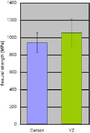 |
| Fig. 1 VITA internal measurements of density to bending in 3 points, Equipment DIN EN ISO 6872. |
Combination of high density and extreme
resistance to fractures of Y-TCP, provide the expectations of endurance
and high clinical rate of successful restorations at permanent loading.
High resistance of Y-TCP to fractures is obtained by so-called
reinforcement of transformation. ZrO2 crystals, present in
Y-TCP, are supplement Y2O3 in metastable
tetragonal phasic condition, which unstable ZrO2 achieves in
the usual way at the temperature ranging from 950 degrees C to 2370
degree C. If now the tetragonal circonium is induced with energy in the
form of a fracture passing through material, then circonium oxide
crystals are converted into thermodynamic more stable monocline phase
with increased volume at room temperature. Energy used on transformation
and tensions under pressure occurring in the structure due to increased
volume, retards or even holds up fractures.
The measure fracture resistance of VITA In-ceram YZ CUBES amounts to 5,9
MPa.vm (SEVNB method, Single-Edge-V-Notched-Beam).
These values allow making the long-span bridges, construction of highly
aesthetic little cross-section connectors as well as carrying out the
preparation that protects dental substance in all-ceramic restorations.
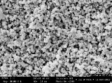
Due to their controlled porous microstructure, VITA in-Ceram YZ CUBES
can be precisely shaped by grinding with SIRONA INLAB apparatus (Fig.
2.). Isotropic shrinkage of material by about 25% in the course of
sintering process brings to the homogenous structure (Fig. 3). It
enables extremely matching restorations and provides high quality
surface.
Crowns D in connection with VITA ceramic, and adequate bonder, with
warmth amplifying coefficient (WAC) perfectly set on Y-TCP, produce
complete aesthetic ceramic restoration.
 |
 |
| Fig. 2 Microstructure (Controlled porosity of sintering) of VITA In-Ceram YZ CUBES for CAD/CAM grinding. | Fig. 3 Microstructure of densely sintered VITA In-Ceram YZ CUBES ceramics after CAD/CAM grinding |
Investigation of Matching Accuracy
The investigation (Le Tran 2003) carried out in vitro at the Laboratory of Computer Aided Dental Restorations showed very good matching in relation to control method as showed in Table I
Table1. Marginal matching accuracy of bridge crowns on
abutments
Marginal notch width (µm) of the tripartite bridge abutment crownworks (X± SD; n=6) at preparation in procedure In-Ceram TZ Cubes (VITA) / CERES InLab (SIRONA and in procedure CERCON (DEGUSSA) in staircase and notch construction. Control period (min) per construction (X±SD; n=6; Le tran 2003
|
Marginal matching accuracy of bridge crowns on abutments |
|||
|
Procedure |
Preparation |
Notch width(µm) |
Monitoring (min) |
|
CEREC/In-Ceram YZ |
Shoulder |
52,9±20 *** |
15,8±1,1 |
|
CEREC/In CeramYZ |
Chamber |
71,1±34 * |
16,3±0,8 |
|
CERCON |
Shoulder |
119,9±34 |
30,1±2,3 |
|
CERCON |
Chamber |
124,1±57 |
32,0±2,2 |
![]() *P<0,05; ***P<0,001;
t-Test
*P<0,05; ***P<0,001;
t-Test
![]() Clinical case
Clinical case
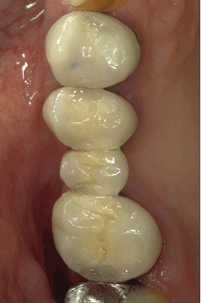
A 58 year-old patient wanted to replace a metal ceramic crown on tooth 14 (VMC-crown) and metal-ceramic bridge (VMC-bridge) on teeth 15-17 with all-ceramic reconstruction without metal (Fig 4a). The abutment teeth were devitalized.
Preparation and Modeling
After removal of VMC constructions, old superstructures were replaced with adhesive composite superstructures (Tetric, Ivoclar, Vivadent) and staircase preparation was applied (Fig. 4b). The stair width was 1 mm. Pronounced notched preparations with clearly recognizable edge shaping were also done. Prior to modeling, retracting strands were put (Ultrapac, Ultradent) into gingival suclus. Dental casts were taken twice using Permadin (3M ESPE). Upper jaw position was registered arbitrary according to facial arch (Artex, Girrbach) and transferred on articulator (Artex).
|
|
|
|
|
|
Fabrication of construction, trial, blending (Shading)
Cross section model was made and articulated with lower jaw central bite at the dental technical laboratory. Additionally, scan model in plaster cast was made, and matted white with scan spray (Dentaco). The dental technician mounted scan-model on the scan-holder CEREC Inlab apparatus (Sirona) (Fig 5). The abutment axis directions were positioned as vertical as possible on the axis spindle. Then, the scan process was started. The technician made the bridge construction in the ready digital 3D-scan-image, drawing manually only the preparation borders of abutments and entered the thickness of crownworks walls (0,6 mm). Intermediate element and connectors were shaped by simple selection of parameters listed in the menu.
The dental technician selected “VITA in-Ceram YZ Cubes” in the menu of materials and placed “VITA in-Ceram YZ Cubes” in the grinding chamber. When calculating the dimensions of the working model, the program has automatically enlarged the bridge construction by 24% to compensate shrinkage at later sintering, and INLAB apparats (Sirona) grinded the bridge construction in accordance with enlargement. The ceramic was sintered into dense structure using specialized furnace (Vita) (Fig. 3).
After the removal of temporary bridge, the matched bridge construction was mounted on cleaned tooth abutments and marginal matching was checked. Thermoplastic “Impression Compound” (Kerr) was spread over the construction and center was registered. Shading color selection as well as shading ceramic (Vita D) selection was made by the dental technician.
|
|
|
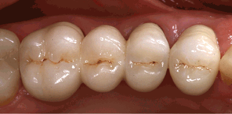
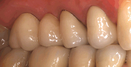
| Fig. 5 Construction of crowns and bridge on model. | Fig. 6 Vitadur D used for shading (coloring) of tripartite bridge YZ Cubes from 15 to 17 and premolar crown of the construction on 14 |
Final procedure
The dentist tried the shaded bridge, and
checked matching, aproximal contacts and occlusion. Prior to adhesive
insertion, inner surfaces of crown holders were treated with aluminum
oxide (50 Im, 3-4 bars).
The bridge and crown were inserted adhesively with Panavijom 21 TC
(Kuraray). The dental nurse mixed the material according to
instructions of manufacturer and spread it on the lumen of polished
working crowns evenly with a brush. By gentle pressure, the bridge was
mounted on the tooth roots previously treated with Syntac Classic
System (Ivoclar Vivadent). The dentist removed sufficient
material with a probe carefully, not to pull the material out of
notches. The aproximal contacts were checked with dental silk. Then, the
isolating gel (Oxyguard II, Kuraray) was put into the area of
notch to avoid inhibitory layer of oxygen on the surface. The time of
Panavia 21 TC binding amounts to 7 min. The dentist removed the
excesses and blew off the isolating gel. At the end, once again the
functional control was carried out. Adhesive insertion seems simple, and
according to our experience, provides better aesthetic results and
marginal sealing in comparison to classic cements. 1,2 (Fig.
6),
Literature
1. Bindl A., Mörmann W.H. : An up to
5-year clinical evaluation of posterior In-Ceram CAD/CAM core crowns.
Int. J Prosthodont. 2002;15:451-456.
2. Mörmann W.H , Bindl A : All-ceramic chair-side computer-aided
design/computer-aided machining restorations. Dent Clin N Am. 2002
;46:405-426.
A. Bindl
Tooth Color Restoration
and Computer Aided Res-toralion Station of the Preventive Dental
Medicine,
Periodontology and Cariology Clinic, Zurich University Center for
Dental,
Mouth and Jaw Diseases, Plaltenstr. 11,
8028 Zurich, Svvitzerland.
e-mail:
sekretariatszcr@zzmk.unizh.ch
- Copyright © 2003. by The Editorial Council of The Acta Stomatologica Naissi
