Introduction
The entire excision of cancer including the adjacent healthy tissue is
one of the basic principles in oncologic surgery. In the lower lip
cancer surgery, the excision of the minimum 1 cm of healthy tissue has
become a standard principle1. For that reason even small
cancers (up to 1cm in diameter), create postoperative lip defect larger
than 3 cm. It is clear that the functional lip reconstruction is
problematic if the postoperative defects exceed 50%, especially when
there is a need to meet the basic requirements of lip reconstruction,
that is to provide the newly formed lip the same or about the same as
the natural lip. Every reconstructive intervention should meet the following
principles: provision of lip support and position, lip contour
restoration, adequate lip sulcus and mouth opening, and maintenance of
senssation2. In clinical practice, the principle of simple
reconstruction of full lip thickness after cancer ablation is frequently
applied in cases of up to 30%-diameter defects of the lip width. In such
cases, V or Y cancer excision is frequently applied and lip defect
is reconstructed by simple approximation of lip ends. Yet, the
prevailing approaches suggest that the radicality must not be abandoned
despite the size of lost lip tissue, for lips can be successfully
reconstructed by flaps even in defects of lager dimensions3.
This study presents the surgical technique of functional reconstruction
of lower lip with 60%-postoperative defect of the lip width. The mental
V-Y island neurovascular muscle-cutaneous flap is applied. This flap,
i.e. technique was introduced in surgical practice in 1997 by Bayramicli
et al.4. In our clinic, this technique has been applied since
1998.
Surgical
technique
Firstly, the tumor limits are defined including at least
1cm of healthy tissue surrounding the cancer. After that, the
topographic position of the mental foramen that is mental arteria and
nerv an "X" is marked on the chin skin. Methylene blue is used for
drawing the flap in the shape of a triangle (V-Y) so that the caudal
point of the flap is placed underneath mandible; neurovascular mental
bundle is a constituent part of the flap and it is preferable to be in
the middle of the flap. The defect of rectangular shape remains after
the cancer excision. Then, the marked skin is notched to the subdermal
tissue. After deepening the cut to the periosteum in the medial part,
the flap elevation starts from medial toward the lateral part. It is
necessary to prepare the tissue carefully, and to identify the mental
neurovascular bundle. Then, the joint of the depressor lip muscles,
composing parts of the flap, is separated on the mandible basis. The
next incision in the lateral part of the flap (it is necessary to
preserve the remnants of m.orbicilaris oris, m.depressor anguli oris and
/or m.depressor labii inferioris) enables the flap mobilization after
the separation of muscle joints from the mandible. After incisions and
mobilization of the flap, it is possible to move the flap by advancement
into the postoperative defect. It is possible to use local mucosal flaps
if there is a defect of the mucosa on the oral side of the flap.
Reconstruction of the oral sphincter is done with the remnants of
m.orbicularis oris or, if it is excised, by reorientation of the fibers
of m. depressor anguli oris or m.depressor labii inferioris. The skin
and muscles are saturated separately in two layers thus forming the V-Y
island muscle-cutaneous neurovascularized advancement flap (Scheme 1).
|
|
|
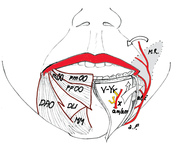
Scheme 1. Surgical anatomy of the lower lip. OO-m.orbicularis oris; pmOO-pars marginalis m.orbicularis oris; ppOO-pars peripheralis m.orbicularis oris; DAO-m.depressor anguli oris; DLI-m.depressor labii inferioris; MM-m.mentalis; a.m./n.m-a.mentalis and n.mentalis; a.f.-a.facialis; a.l.i.-a.labialis inferior; MF-mucosal flap; V-Yr-mental V-Y island neurovascular advancement flap. |
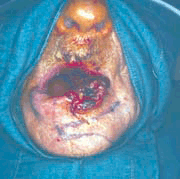
In male patient J.M., aged 84 years, bleeding planocellular lower lip
cancer was diagnosed. Patient J.M. had pathologically altered internal
status, and laboratory blood analysis were considerably exceeding the
upper limit values. For that reason, the general endotracheal anesthesia
was not applicable, but surgery under local anesthesia was planned. The
cancer was located from the lip median line to the left lip commissure,
and included left lateral pericommissural lip tissue on the left side as
well. Horizontally, the cancer was 32 mm in diameter, and vertically 22
mm (Fig. 1). Before the surgery began, bilateral conductive anesthesias
for n.mentalis and left sided for n.buccalis were administrated, as well
as 5 ml of the 2% anesthetic Lidokain®-Adrenalin (ICN Galenika,
Belgrade). After designing the cancer edges (including minimum 1 cm of
the surrounding healthy tissue) and V-Y island muscle-cutaneous flap,
lip cancer was excised (Fig. 2). Postoperative defect amounted to 52 mm
horizontally, and 22 mm vertically. Defect extended laterally, toward
healthy part of the lip, from the median line to the cheek in the area
of lip mediolus and vertically, to sulcus mentolabialis. Careful flap
mobilization was conducted along with the identification of
neurovascular mental bundle and depressor anguli oris muscle laterally
(Fig. 3). Then, the mobilization of entire flap was conducted cranially
for restoration of postoperative defect. The flap was saturated in two
layers by atraumatic technique. The mucosa and muscles were saturated
with resorptive strand Vicryl 5-0 (Ethicon, Edinburgh, Great Britain),
and the skin with atraumatic silk strand Perma-Hand® Seide 4-0 (Ethicon,
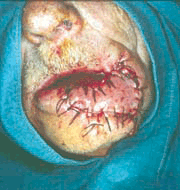
Nordestedt D-22851, Germany). The vermilion was reconstructed by the
mucosal flap, and suturated with the same strand as the skin (Fig. 4).
Postoperatively, the patient received equalized antibiotic therapy of
Lincocin® 600 mg / 2ml i.m every 12 hours (Hemofarm D.D., Vrsac) for 7
days. The patient was discharged in good condition seven days after the
surgery, and the flap vitality was complete and with no signs of
infection and necrosis (Fig. 5).
 |
 |
 |
 |
 |
|
Figure 1. Bleeding planocellular lower lip cancer in exophthytic form.
|
Figure 2. Marking of incision lines 1cm entering the adjacent healthy tissue with prior topographic orientation of the mental opening in the flap derma.
|
Figure 3. Flap elevation with identification of mental arteria and nerv (marked with arrow).
|
Figure 4. Intraoperative appearance of reconstructed lip
|
Figure 5. Postoperative appearance of reconstructed lip V-Yr seven days after surgery. The flap is completely vital, and the reconstruction of pericommissural tissue is conducted so that newly formed lip almost completely corresponds to the natural one. |
Discussion
The most reliable way of lower lip cancer treatment is the surgical excision. Postoncological reconstruction of the lower lip is supposed to provide the newly formed lip as functional as the normal one. Defects of 30-60% of the lip size, require the use of flaps what, as it should be known, in most techniques compromises the innervation, vascularization, or does not provide the reorientation of muscle fibers 5-7. Functional lip reconstruction is possible if the Karapandzic technique of arterialized neurovascular flap elevation is applied 8. This technique, considered to be almost ideal, enables the reconstruction of defects ranging from 30-80%. The main imperfection of Karapandzic technique is the postoperative microstoma already in lip defects of 60%. Additionaly, using of Karapandzic flap is not reliable in lower lip reconstruction when the blood vessels are lost by neck dissection (a.facialis). By application of mental V-Y island neurovascular muscle-cutaneous flap, postoperative microstoma is avoided, and flap innervation is completely preserved since n.mentalis is preserved. The risk of ischemia and necrosis of the mental V-Y island neurovascular muscle-cutaneous flap is avoided, for this flap is provided with double and independent arterial nourishment: by a. labialis (branch of a.facialis) and a. mentalis (a.alveolaris inferior). According to vascular anatomy, the mental V-Y island neurovascular muscle-cutaneous flap of the lip is classified, along with the muscles (m.orbicularis oris and m.depressor anguli oris and/or m.depressor labii inferioris), as type III muscle-cutaneous flap. Type III flaps have two independent vascular pedicles 8. This makes the surgeon free to conduct necessary neck dissection, if necessary without preserving a.facialis, and than to conduct a lip reconstruction freely, using the flap with preserved vascularization. In lip reconstruction it is necessary to place the lip commissure in the same plane with the opposite one. However, the imperfections in the symmetry of new lip, as well as the formation of postoperative macrostoma are frequent defects of particular techniques such as McGregors6 or Fries5 or additional incisions, localized out of anatomical cancer localization (e.g. in the check), are needed for flap mobilization; such procedure has a negative aesthetic component10. By application of the mental V-Y island neurovascular muscle-cutaneous flap, macrostoma is not formed for it is possible to measure the accurate amount of removed tissue and to move by advancement the same amount of tissue into the postoperative defect. Additionally, flap positioning in the area of commissure is possible and easy, since after the proper elevation technique, the flap is sufficiently mobile for accomplishing the symmetry of lip commissures. The incisions for elevation of the mental V-Y island neurovascular muscle-cutaneous flap are within anatomical borders proving favorable aesthetic outcome. It is particularly important to point to the fact that the reconstruction of the orbicular sphincter is conducted with the remnants of m.orbicularis oris, m.depressor anguli oris and m.depressor labii inferioris. With these muscles, isolated or grouped, it is possible to obtain the motor function of reconstructed lip as the n.facial ending motor branches innervating the mentioned muscles are preserved. In addition, the reconstruction of lip mucosal part is easy because there are mucosal flaps available for reconstruction of the vermilion. The lip reconstructed with application of the mental V-Y island neurovascular muscle-cutaneous flap completely looks like the natural lip in relation to depth, length, height and position, what is noticeable in the presented case. Based on the presented case, we can recommend the application of the mental V-Y island neurovascular advancement flap as one of the alternative techniques in the functional reconstruction of lip defects of 30-60% of its length.
Literature
- Cruse C.W., Radocha R.F.: Squamous cell carcinoma. Plastic and Reconstructive Surgery. 1987;80:787-791.
- Jabaley M.E., Orcutt T.W., Clement R.L.: Application of Karapandić principle of lip reconstruction after excision of lip cancer. Am. J. Surg. 1976;132:529-532.
- Jackson I.T.: Lip reconstruction. (In) Local flaps in head and neck reconstruction. St.Louis; C.V. Mosby, 1985.
- Bayramiçli M., Numanov A., Tezel E.: The mental V-Y island advancement flap in functional lower lip reconstruction. Plastic and Reconstructive Surgery. 1997; 100:1682-1690.
- Fries R.: Advantages of a basic concept in lip reconstruction after tumor resection. J. Maxillofac. Surg. 1973; 1:13-18.
- McGregor I.A.: Reconstruction of the lower lip. Br. J. Plast. Surg. 1983;36:40-47.
- Nakajima T., Yoshimura T., Kami T.: Reconstruction of the lower lip with a fan shaped flap based on facial artery. Br.J.Plast.Surg. 1984;37:52-54.
- Karapandić M.: Reconstruction of lip defects by local arterial flaps. Br.J.Plast.Surg.1974;27:93-97.
- Mathes S.J., Nahai F.: Classification of the vascular anatomy of muscles: experimental and clinical correlation. Plastic and Reconstructive Surgery. 1981; 67: 177-187.
- Webster J.P., Coffey R., Kelleher R.E.: Total and partial reconstruction of the lower lip with innervated muscle-bearing flaps. Plastic and reconstructive Surgery. 1960; 25:360-371.
Medical Faculty, Clinic of Stomatology
Oral & Maxillofacial Surgery,
52 Braće Tasković str.
18000 Ni
Serbia (Serbia and Montenegro)
E-mail: nburic@yahoo.com.
- Copyright © 2003 by The Editorial Council of The Acta Stomatologica Naissi