IIntroduction
Diseases of the periodontium (gingivae, cement, alveolar bone and periodontium) are frequent, presenting in children as well as in adults. The etiology of these diseases is complex; clinical manifestations are different making the classification more difficult. One classification, presented in 1999, foremost is based on the etiology of these diseases.1,2
CLASSIFICATION OF PERIODONTAL DISEASES 1
I GINGIVAL DISEASES
A: Dental plaque-induced gingival diseases
1. Gingivitis associated with dental plaque only
2. Gingival diseases modified by sistemic factors
a) Associated with the endocrine system
1. Puberty associated gingivitis
2. Menstrual cycle-associated gingivits
3. Pregnancy associated
4. Diabetes mellitus- associated gingivits
b) Associated with blood dyscrasias
1. Leukaemia-associated gingivitis
2.Other
3. Gingival diseases modified by medications
a) Drug-influenced gingival diseases
1. Drug-influenced gingival enlargements
2. Drug-influenced gingivitis
a) Oral conraceptive-associated gingivitis
b) Other
4. Gingival diseases modified by malnutrition
B. Non-plaque-induced gingival lesions
1. Gingival diseases of specific bacterial origin
2. Gingival diseases of viral origin
a) Herpes virus infections
1. Primary herpetic gingivostomatitis
2. Recurrent oral herpes
3. Varicella-zoster infections.
4. Other
3. Gingival diseases of fungal origin
a) Candida species infections
b) Linear gingival erythema
c) Histoplasmosis
d) Other
4. Gingival lesions of genetic origin
a) Hereditary gingival fibromatosis
b) Other
5. Gingival manifestations of systemic con-ditions
a) Mucocutaneous disorders
1. Lichen planus
2. Pemphigoid
3. Pemphigus vulgaris
4. Erythema multiforme
5. Lupus erythematosus
6. Drug-induced
7. Other
b) Allergic reactions
1. Dental restorative materials
2. Reactions attributable to
a) Toothpastes/dentifrices
b) Mouthrinses/mouthwashes
c) Chewing gum additives
d) Foods and additives
3. Other
6. Traumatic lesions
a) Cemical injury
b) Physical injury
c) Thermal injury
II CHRONIC PERIODONTITIS
A) Localized
B) Generalized
III AGGRESSIVE PERIODONTITIS
A) Localized and generalized periodontitis
B) Rapid periodontitis
C) Refractory periodontitis
IV PERIODONTITIS AS A MANIFESTATION OF SYSTEMIC DISEASES
A) Associated with haematological disorders
1. Acquired neutropenia
2. Leukaemias
3. Other
B) Associated with genetic disorders
1. Familial and cyclic neutropenia
2. Down syndrome
3. Leukocyte adhesion deficiency
syndromes
4. Papillon-Lefevres syndrome
5. Chediak-Higashi syndrome
6. Histiocytosis syndrome
7. Glycogen storage
disease
8. Infantile genetic
agranulocytosis
9. Cohen syndrome
10. Ehlers-Danlos syndrome (Types IV and VIII)
11. Hypophosphatasia
12. Other
V NECROTIZING PERIODONTAL DISEASES
A) Necrotizing ulcerative gingivitis
B) Necrotizing ulcerative periodontitis
VI ABSCESSES OF THE PERIODONTIUM
A. Gingival abscess
B. Periodontal abscess
C. Pericoronal abscess
VII PERIODONTITIS ASSOCIATED WITH ENDODONTIC LESIONS
A. Combined periodontic-endodontic lesions
VIII DEVELOPMENTAL OR ACQUI-RED DEFORMITIES AND CONDITIONS
A. Localized tooth-related factors that modify or
predispose to plaque-induced gingival diseases/periodontitis
1. Tooth anatomical factors
2. Dental restorations/appliances
3. Root fractures
4. Cervical roort resorption and cemental tears
B. Mucogingival deformities and conditions around
teeth
1. Gingival/soft tissue recession
2. Lack of keratinized gingiva
3. Decreased vestibular deppth
4. Aberrant frenum/muscle position
C. Mucogingival deformities and conditions on edentulous ridges
D. Occlusal trauma
Knowing and early detection of risk factors is
necessary for correct and timely diagnosis as well as for adequate
treatment of those diseases.
According to the modern literature, the risk
factors are:
- Genetic predisposition
- Race
- Gender
- Intercurrent diseases (neutropenia secondary,
diabetes mellitus, acute and chronic leukemia, agranulocytosis,
transient immuno-suppression)
- Drug usage (calcium antagonists, antie-pileptics,
cyclosporins, chloramphenicol, oral contraceptives, antiinflammatory
drugs)
- Nutrition
- Socioeconomic status
- Age
- Smoking
- Osteoporosis
- Poor oral hygiene
- Presence of iatrogenic factors
- Mucogingival anomalies
The treatment of diseases of the perio-dontium is
complex. Therapy plan is strictly individual depending on local and
general status of a patient.1,2
The aim of the therapy is:
- Identification, elimination and control of dental
plaque and other favoring factors
- Elimination of the symptoms of inflamma-tion and
destruction of the periodontal tissue in order to stabilize the tissue
resistance and establish morphological, functional and occlu-sal
relations.
The therapy success depends on the following
factors:
- Patient's motivation and cooperation
- Oral hygiene
- Age
- Patient's occupation
- General health condition
- Duration (course) of the disease
- Condition of the diseased periodontium
- Condition of occlusion and articulation
- Presence of anatomical anomalies
- Morphology of the gingiva and teeth
- Malocclusion
- Functional demands
- Number and position of the teeth
- Condition of the alveoloar bone
- Bad habits
All therapeutic procedures in the treatment of periodontal disease are classified into initial - causal or non-surgical methods and surgical methods.
INITIAL-CAUSAL THERAPY
MOTIVATION AND INSTRUCTION OF THE PATIENT TO FOLLOW NORMAL ORAL HYGIENE PRACTICE
Basic, most significant, but the most difficult task is to motivate the patient to realize the importance of dental plaque and regular and correct tooth brushing. Dental plaque is the enemy of oral health care, and oral hygiene is the enemy of dental plaque.1
SUPRAGINGIVAL AND SUBGINGIVAL PLAQUE ELIMINATION AND CONTROL
Plaque control is conducted:
1. in a mechanical way through home control by the
patient himself using the means for maintenance of oral hygiene; and
professional control by the dentist during follow-ups).
2. by chemical means used as a supplement to the
conventional therapy, and in treatment of some complications and forms
of periodontal disease. They can be nonspecific and specific.
Among nonspecific means the most frequently used
are: chlorhexidine digluconate hibitane. It is used in the form of gel,
solution, emulsion, toothpaste and Periochipe.
Benzocaine chloride, Cetyliridium chloride, Kalium
fluoride and phenol substances proved less effective in decrease of
dental plaque and inflammation of the gingiva.
Among specific chemical agents, antibiotics are
used and administrated systemically or locally, according to
antibiogram.
Antibiotic selection depends on the dominant
microbiologic flora. Antibiotics are used in acute conditions in the
periodontium (ulcero-necrotic changes, periodontal abscesses), and as
adjuvant therapy in juvenile, rapid and refractive periodontal disease,
and before and after surgical treatment of periodontal disease.2
In local application, biocompatible (ethylene wynil
acetate - EVA system), systems are used, acrylic and ethyl cellulose
stripes, viscose gels, bioresorptive capsules and irrigation systems.
The most often used are:
TETRACYCLINE administrated 4 times daily at
therapeutic dose of 250-500 mg up to three weeks
DOXYCYCLINE and MONOCYCLINE a semi synthetic
tetracyclines administrated at a therapeutic dose of 200 mg or 100mg
daily over three weeks.
METRONIDAZOL is a bactericidal chemo-therapeutic,
nitroimidazol chemical compo-sition. It acts on gram+ and Gram-
bacteria. On the market, it can be found under the names Orvagil and
Flagyl. It is applied at a dose of 250-500 mg three times a day over
7-14 days. Nowadays, it is one of the medicines of choice in treatment
of periodontal disease.
CIPROFLOXACIN acts on gram bacilli.
PENICILLIN is administrated in high doses, in the
infections caused by bacillus fuziformin and Vensan spirochete. It is
administrated at a dose of 250-500 mg over 5-10 days.
AMOXYCILLIN is administrated in combi-nation with
clavulanate-acid which inhibits beta lactamines, products of bacteria.
SPIRA-MYCIN AND ERYTHROMYCIN are macrolides acting
on Gram+ bacterium Staphylococcus aureus and Actinomyces viscosus.
CLINDAMYCIN is administrated in a dose of 250 mg
over 7-10 days.
ELIMINATION OF FAVORING FACTORS
During the course of this therapeutic proce-dure, dental calculus and caries, poor conser-vative fillings and prosthetic works, causes of food impaction, bad habits and mucogingival anomalies are eliminated and corrected. 2
SUBGINGIVAL CURETTAGE
Subgingival curettage is a basic procedure in healing the periodontal pockets. Subgingival curettage includes also the procedure of eliminating subgingival concrements and necrotic cement, changed epithelium of the periodontal pocket, granular tissue and inflamed subepithelial connective tissue. This procedure is indicated in periodontal pockets of 4-6mm.1,2
APPLICATION OF PHYSICAL METHODS
Physical methods used in the therapy of diseases of the periodontium are cathode galvanization, drug electrophoresis, igni-punction, periodontal bandages and gingival massage. The usage of lasers (soft and hard lasers) in increased is growing. Soft lasers are applied in treatment of gingivitis, periodontal disease, periodontal abscesses etc, for their anti-inflammatory, anti-edematous, analgesic and biostimulation effect. Hard lasers are used in periodontal surgery.1,2
CONTROL OF GENERAL FACTORS - SYSTEMIC FACTORS
Systemic diseases decrease general immunity, and thus change the local response of the periodontal tissue to microorganism actions, therefore drug administration, nutrition, smoking, intercurrent diseases and other factors should be controlled constantly.1,2
OCCLUSION CORRECTION
In this therapy phase, the correction of rough
occlusive disorders of some teeth, central occlusion, protrusive and
lateral occlusive movements. In this way the occlusion balance is
achieved which reduces the intensity of masticatory forces. Diagnosis of
traumatic occlusion, selective grinding, orthodontic treatment and
temporary splints are made prior to surgery.
After surgery, prosthetic management of the patient
is provided and final splints for dental fixation are made.1,2
SURGICAL THERAPY
Surgical therapy understands radical treatment of
diseases of the periodontium and it is preceded by the initial therapy.
Periodontal surgery includes all surgical
interventions involving soft and hard perio-dontal tissues. 3-6
The aim of these interventions is:
- Better approach to deeper periodontal tissues
(treatment of root and bone surface)
- Elimination of periodontal pockets
- Reconstruction of the gingiva and bone
fun-ctional anatomy to provide optimal conditions for dental plaque
control.
Division of periodontal surgery
Gingival and periodontal surgery
- Gingivetomy
- Gingivoplasty
Mucogingival periodontal surgery
Flap
Free mucogingival autotransplant
Frenectomy
Edlan-Mejchar's surgery
Bone periodontal surgery
Resectional
Additional application of bone
transplants and implants
Regenerative guided tissue
regeneration
GINGIVECTOMY WITH GINGIVOPLASTY
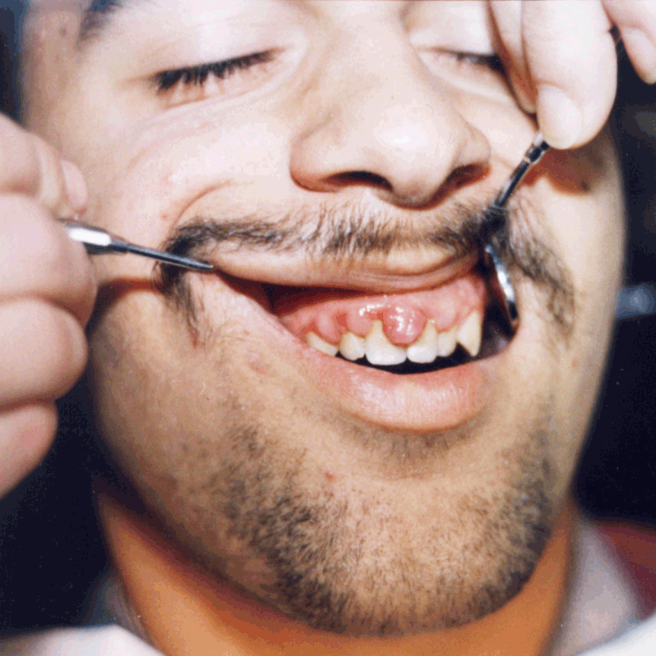
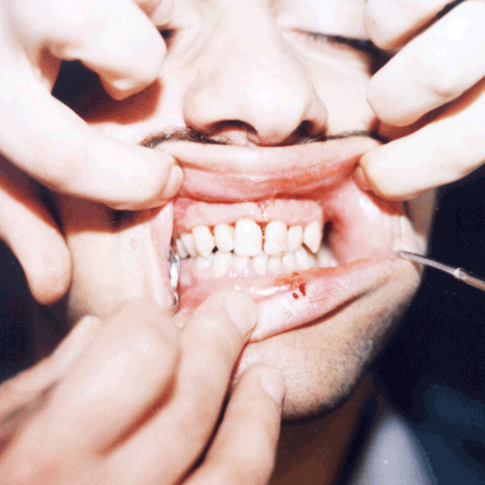
This surgical procedure serves for the removal of periodontal pocket soft tissue and correction of appearance and function of the gingiva. ( Fig 1,2)
It is indicated in the proliferation of connecti-ve tissue, gingival fibromatosis, suprabone periodontal pockets, preprosthetic elongation of the clinical crown and gingival correction procedures.2-4
FLAP SURGERY
Kuster and Partsch 1900
Neuman 1911
Leonard Widman 1916
Ramfjord 1973 modified Widman
flap
surgery
With regard to depth, the flap can be
-Semi-thick flap - mucosal (epithelium and part of
connective tissue)
-Full-thickness flap mucosa + periost
According to position, the flap can be:
1. repositioned (shifted) modified Widman flap
2. positioned -after elevation, the flap is
positioned into new, different positions:
- apically positioned flap
- coronary repositioned flap
- bipedicle flap
INDICATIONS FOR FLAP SURGERY
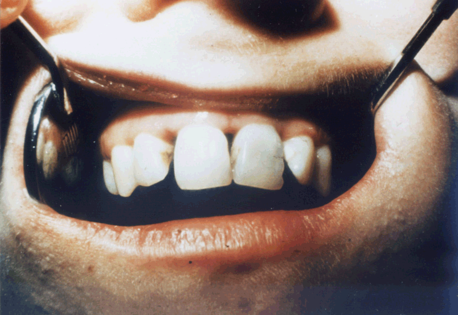
1. Treatment of deep suprabone and infra-bone
periodontal pockets ( Fig 3, 4)
2. Insertion of implants into infrabone
perio-dontal pockets and other transplants
3. hemisection, amputation and bisection of tooth
root
4. Treatment of periodontal abscesses
5. Preprosthetic preparation of the patient
APICALLY REPOSITIONED FLAP
(Naber and Fridman, 1957)
The flap is repositioned apically in relation to
initial position of the flap.
INDICATIONS:
- Elimination of periodontal pockets of su-prabone
and infrabone type reaching mucogin-gival line, or surpassing it.
- Extension of attached gingiva
- Deepening of the vestibule
- Repositioning of frenulum and lateral papi-llae
junction2.
CORONARY REPOSITIONED FLAP
The flap is moved in the coronary direction aiming at elimination of periodontal pockets and accomplishment of attached gingiva to exposed root surface.1-3
LATERALLY REPOSITIONED FLAP
Technique of laterally dislocated flap is used in the case of exposed tooth root, especially in the front teeth due to the gingival withdrawal/detachment/insulated. The exposed tooth root is covered by the flap from the region of adjacent teeth.1-3
DOUBLE PAPILLA FLAP
It is used for covering the exposed tooth root surface. It is applied when the periodontium tissue from neither distal nor mezial side of the exposed root is suitable for lateral flap movement.1-3
FREE MUCOGINGIVAL AUTOTRANSPLANTANT (FMAT)
This method of mucosa repositioning from one place to another is introduced by Bjorn in 1963.
INDICATIONS:
- Formation or extension of attached gingiva
- Elimination of coronary joining frenulum of lips,
tongue and lateral mucosa plicae
- Prevention and treatment of recesion of the
gingiva and covering the exposed roots
- Deepening of fornix4
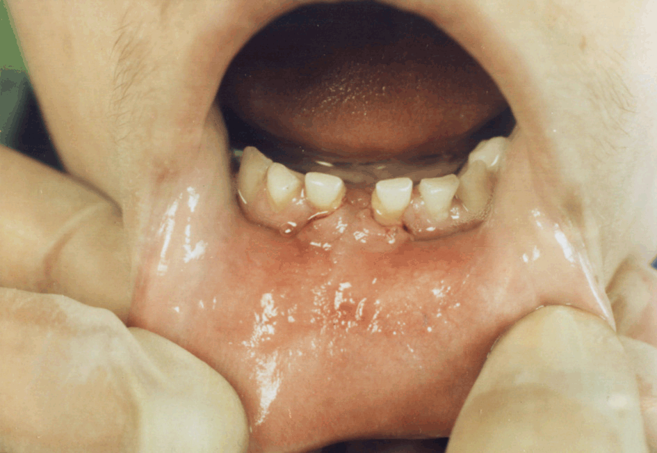
Frenectomy
Frenectomy is a surgical intervention by which too coronary inserted frenula of lips, tongue and lateral mucose plicae are fully eliminated. It is indicated for orthodontic reasons, preventive and therapeutic reasons in diseases of the periodontium and preprosthetic preparation for the purpose of better prosthesis retention.2-4 ( Fig 5,6).
EDLAN-MEJCHAR SURGERY
It is used for deepening the vestibule. Nowadays it
is used less frequently.
In the clinical practice, some modifications of
this method are used.2
RESECTIONAL BONE SURGERY
The gingival profile is in direct relationship with
deeper periodontal tissues, that is with the bone structure.
Diseases of the periodontium lead to bone
re-sorption changing the bone architecture and posi-tioning the bone
more apically to the tooth root.
With resectional surgery bone defects are
eliminated, and so called physiological architecture of the alveolar
bone is created but at a more apical level.5
Therefore, the INDICATIONS are:
- bone remodeling
- elimination on bone craters and bone angle
defects, and
- reconstruction of physiological profile
OSTEOPLASTY
(Fridman, 1955)
The aim of this technique is bone remodeling
without elimination of bone tissue for the purpose of achieving
physiological form.2-4
OSTEOCTOMY
This term understands the elimination of supportive
bone to achieve physiological profile of the bone ridge. It is indicated
in the presence of bone craters or defects, and usually vestibular or
oral part of a bone is removed.2-4
TOOTH RESECTION
If the pathological process compromises the bone
tissue in furcations, the surgical treatment is complex. To eliminate
the periodontal pocket, it is usually necessary to modify the tooth
anatomically, or to eliminate a part of the tooth with one or more
roots.2-4
a)TOOTH HEMISECTION
It is conducted in the molars, mostly lower. This
intervention includes the separation of a tooth in the axial direction,
and afterwards extra-ction of one half of the tooth (crown partly and
root) of which the periodontium is destroyed. On the cut part of the
tooth, prosthetic constru-ction is made up after endodontic
treatment.2-4
b) TOOTH BISECTION
That is a surgical procedure where two pre-molars
are made out of one. Detachment of the tooth, the approach to the tooth
furcation is pro-vided and eradication of the periodontal pocket.
It is indicated if the periodontium surrounding
both roots is preserved, that is if the destruction involved only the
coronary part of furcation. After endodontic treatment and bisection,
cut tooth halves also should be prosthetically managed.2-4
c) TOOTH AMPUTATION
Tooth root amputation understands cutting one of
the multiroot tooth surrounded with destroyed periodontium.
INDICATIONS:
- Removal of the distal or mesial root of the lower
molars
- Removal of the palatine root and preservation of
the mesio and distovestibular root of the upper molars
- Removal of the mesiovestibular root and
preservation of distovestibular and palatine root
- Removal of the distovestibular root and
preservation of the mesiovestibular and palatine root.2-4
d) TOOTH REMOVAL
Tooth removal is used in elimination of the periodontal pocket when the periodontium is so destroyed that it cannot maintain in the jaw.2-4
TUNNEL PREPARATION
Tunnel preparation understands the preparation of
the alveolar bone and adaptation of soft tissues in the region of
furcation providing the communication in the bucolingual direction
through the furcation involved region. This provides adequate
preservation of the oral hygiene.
“Tunnel preparation” demands no endo-dontic or fix
prosthetic works. Exposed surfaces of the tooth root can be involved by
caries so this method should be applied in persons with excellent oral
hygiene and low caries index.2-4
ADDITIONAL PERIODONTAL SURGERY
In the healing of the periodontal pockets and tis-sue regeneration, transplants and implants are used.
TRANSPLANTS:
- Intraoral and extraoral transplants
- Homeotransplants
- Heterotransplants
- Substituents of the transplant bone
Transplantations are not considered to be very
significant in periodontal surgery.3,5,6
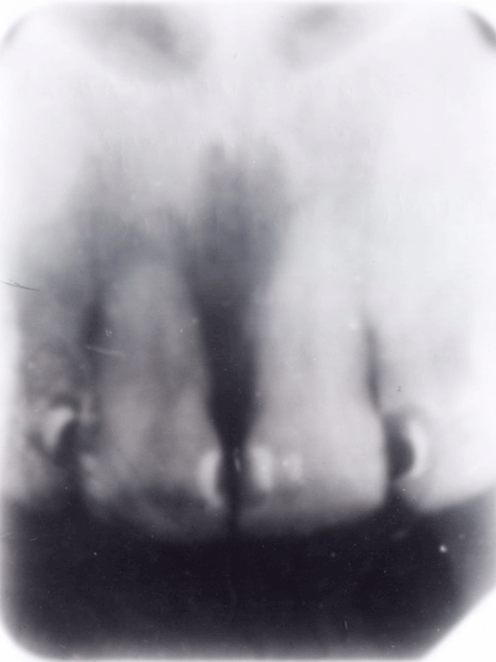
IMPLANTS
Those are synthetic materials for bone replacement
and they make up the destroyed bone, and at the same time induce new
bone formation. Artificial bone has to be: bioinert, compatible with
adjacent tissues, and without antigenic, mutagenic and cancerous
potential.
They are found in granular, grainy and rodlike
shape. They are composed of mineral salts (calcium, phosphorus,
magnesium, alumi-num). Implants should possess oseoinductive and
osteopromotive capacities, that is artificial bone should become a
constituent part of the jaw bone, but to stimulate new bone formation as
well ( Fig 7, 8).
 |
 |
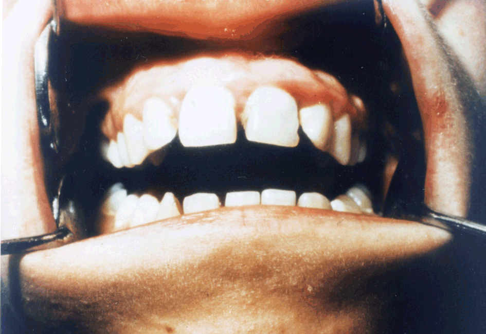 |
| Figure 1. Gingivectomy, before surgery | Figure 2. Gingivectomy, after surgery | Figure 3. Flap operation, before surgery |
 |
 |
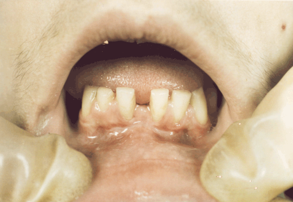 |
| Figure 4. Flap operation after surgery | Figure 5. Frenectomy, with vestibuloplasty - before operation | Figure 6. Frenectomy with vestibuloplasty - after operation |
 |
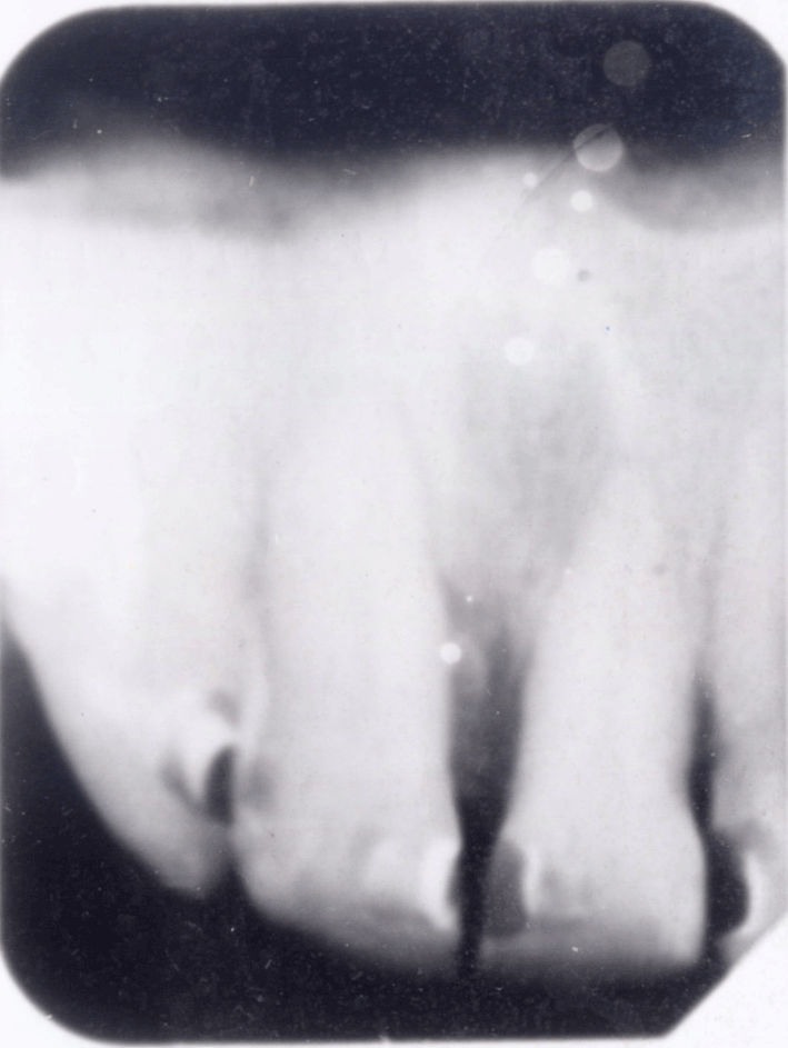 |
| Figure 7. Using of implants, before operation | Figure 8. Using of implants, after operation |
On the market, there is a great number of good
quality materials used for filling the bone defects:
Syntograft, Ceros 80, Ceros 82, Periograf,
Osprovit, Durapatit, Frialit, Calcitite, Alveograf, Allotropat 50,
Interpore 200, Algipore, Biobase, Bio-oss, AlloGro, Biogran, Pep Gen
P-15 as well as domestic preparation Aldovit. Among new materials, the
best known are Osteogenin, Amelogenin, Enamelin, Amlein, Endogein. For
the guided tissue regeneration, the growth factors such as Emdogain with
Pref-gel are added.
INDICATIONS:
- Bone destruction with formation of infra-bone
defects in the jaw bone
- Cysts
- Granulomas
- Injuries
GUIDED TISSUE REGENERATION
In cicatrization of surgical wounds, most
frequently, long epithelial junction is formed with absence of
spontaneous regeneration of bone tissue, periodontal ligament and root
cement.
The cells of epithelial origin are very fast in
migrating apically, covering inner flap surface disabling the joining of
periodontal fibers and tooth root surface. The cells of adhering origin
proliferate more slowly than the epithelial cells and in contact with
tooth root surface without cement they lead to the root resorption.
The cells of bone origin are the slowest in
development. In contact with root surface without cement, periodontal
fibers bring to the root resorption and ankylosis.
The cells of periodontal ligament are the only
cells which possess the capacity of differe-ntiation and creation of
cement, bone tissue and periodontal fibers.
The procedure used in regeneration of the
periodontal tissue lost during the periodontal disease, is guided tissue
regeneration. It is based on the application of membrane barrier
so-called shower curtain effect which isolates the periodontal defect
from the gingival epithelium and connective tissue enabling the renewed
periodontal ligament adherence to the tooth surface. Nonresorptive,
resorptive and perio-steal membranes are used.
The first membranes used were nonresor-ptive (since
1982) such as Polytetra-fluore-thylene (PTFE). Today, the following
resorptive membranes are used: Bio-Guide, Bio-Mend, Allo-Derm, Atrisorb
etc.6
Conclusion
Generally accepted rule is that the remissions can
never be so stable that exclude recurrences. It is considered that one
of the most significant factors of stable remission and recurrence
prevention is the patient cooperation.
The patient has to be instructed to proper oral
hygiene practice, and than, motivated to follow strictly the
instructions, not only in the period immediately following the treatment
but conti-nuing through life.
-
Laskaris G, Scully C. Periodontal Manifestations at Local and Systemic Disease. Colour Atlas and Text. Springer-Springe 2002.
-
Zelić O. Osnovi klini~ke parodontologije, Beo-grad, 2000.
-
Backer W, Backer B. A prospective multicenter study evaluating periodontal regeneration for class II furcation invasion and intrabony deffects after treatment with a bioabsorable barrier membrane: one year results. J Periodontol 1996; 66: 641-649.
-
Kojović D, Martinović S, Branković V: Hiruške korekcije mukogingivalnih odstupanja. Zbornik radova VI kongresa stomatologa Makedonije, 1991, Dojran.
-
Trombelli L, Scabbia A, Tatakis D, Calina G. Subpedicle Connective Tissue Graft Vercus Guided Tissue Regeneration with Bioabsorable Membrane in the Treatment of Human Gingival Recession Defects. J Periodontol 1998; 69:1271-1277.
-
Tinti C, Vincenti G, Cocchettox. Guided tissue regeneration in mucogingival surgery. J Periodontol 1993;64:1184-1191.
Adress for correspondence:
Prof. Draginja Kojović DDS, MSD, PhD
Clinic of Dentistry -Nis
Bra}e Tasković 52 Street
Serbia and Montenegro
- Copyright © 2003 by The Editorial Council of The Acta Stomatologica Naissi